{"title":"Morphology and histology of the olfactory organ of two African lungfishes, Protopterus amphibius and P. dolloi (Lepidosirenidae, Dipnoi)","authors":"Hyun Tae Kim, Jong Young Park","doi":"10.1186/s42649-021-00054-x","DOIUrl":null,"url":null,"abstract":"<p>The olfactory organs of two African lungfishes, <i>Protopterus amphibius</i> and <i>P. dolloi</i>, were investigated using a stereo microscope and a compound light microscope and were described anatomically, histologically, and histochemically. Like other lungfishes, these species present the following general features: i) elongated olfactory chamber (OC), ii) anterior nostril at the ventral tip of the upper lip, iii) posterior nostril on the palate of the oral cavity, iv) lamellae with multiple cell types such as olfactory receptor neurons, supporting cells, basal cells, lymphatic cells, and mucous cells (MC), and vi) vomero-like epithelial crypt (VEC) made of glandular epithelium (GE) and crypt sensory epithelium. Some of these features exhibit differences between species: MCs are abundant in both the lamellar and inner walls of the OC in <i>P. amphibius</i> but occur only in lamellae in <i>P. dolloi</i>. On the other hand, some between feature differences are consistent across species: the GE of both <i>P. amphibius</i> and <i>P. dolloi</i> is strongly positive for Alcian blue (pH?2.5)-periodic acid Schiff (deep violet coloration), and positive with hematoxylin and eosin and with Masson’s trichrome (reddish-brown staining), unlike the MCs of the two species which stain dark red with both Alcian blue (pH?2.5)-periodic acid Schiff and Masson’s trichrome but respond faintly to hematoxylin and eosin. The differing abundance of MCs in the two lungfishes might reflect different degrees in aerial exposure of the olfactory organ, while the neutral and acid mucopolysaccharide-containing VEC, as indicated by staining properties of the MCs, is evolutionary evidence that <i>P. amphibius</i> and <i>P. dolloi</i> are the closest living relatives to tetrapods, at least in the order Dipnoi.</p>","PeriodicalId":470,"journal":{"name":"Applied Microscopy","volume":"51 1","pages":""},"PeriodicalIF":0.0000,"publicationDate":"2021-04-17","publicationTypes":"Journal Article","fieldsOfStudy":null,"isOpenAccess":false,"openAccessPdf":"https://sci-hub-pdf.com/10.1186/s42649-021-00054-x","citationCount":"4","resultStr":null,"platform":"Semanticscholar","paperid":null,"PeriodicalName":"Applied Microscopy","FirstCategoryId":"1085","ListUrlMain":"https://link.springer.com/article/10.1186/s42649-021-00054-x","RegionNum":0,"RegionCategory":null,"ArticlePicture":[],"TitleCN":null,"AbstractTextCN":null,"PMCID":null,"EPubDate":"","PubModel":"","JCR":"Q3","JCRName":"Immunology and Microbiology","Score":null,"Total":0}
引用次数: 4
Abstract
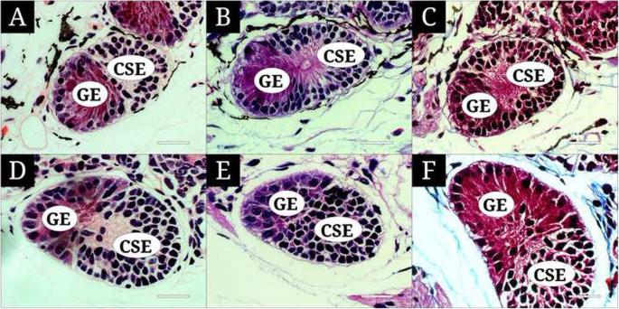
The olfactory organs of two African lungfishes, Protopterus amphibius and P. dolloi, were investigated using a stereo microscope and a compound light microscope and were described anatomically, histologically, and histochemically. Like other lungfishes, these species present the following general features: i) elongated olfactory chamber (OC), ii) anterior nostril at the ventral tip of the upper lip, iii) posterior nostril on the palate of the oral cavity, iv) lamellae with multiple cell types such as olfactory receptor neurons, supporting cells, basal cells, lymphatic cells, and mucous cells (MC), and vi) vomero-like epithelial crypt (VEC) made of glandular epithelium (GE) and crypt sensory epithelium. Some of these features exhibit differences between species: MCs are abundant in both the lamellar and inner walls of the OC in P. amphibius but occur only in lamellae in P. dolloi. On the other hand, some between feature differences are consistent across species: the GE of both P. amphibius and P. dolloi is strongly positive for Alcian blue (pH?2.5)-periodic acid Schiff (deep violet coloration), and positive with hematoxylin and eosin and with Masson’s trichrome (reddish-brown staining), unlike the MCs of the two species which stain dark red with both Alcian blue (pH?2.5)-periodic acid Schiff and Masson’s trichrome but respond faintly to hematoxylin and eosin. The differing abundance of MCs in the two lungfishes might reflect different degrees in aerial exposure of the olfactory organ, while the neutral and acid mucopolysaccharide-containing VEC, as indicated by staining properties of the MCs, is evolutionary evidence that P. amphibius and P. dolloi are the closest living relatives to tetrapods, at least in the order Dipnoi.
Applied MicroscopyImmunology and Microbiology-Applied Microbiology and Biotechnology
CiteScore
3.40
自引率
0.00%
发文量
10
审稿时长
10 weeks
期刊介绍:
Applied Microscopy is a peer-reviewed journal sponsored by the Korean Society of Microscopy. The journal covers all the interdisciplinary fields of technological developments in new microscopy methods and instrumentation and their applications to biological or materials science for determining structure and chemistry. ISSN: 22875123, 22874445.


 分享
分享
 求助内容:
求助内容: 应助结果提醒方式:
 扫码关注我们
