{"title":"Pore Formation by Amphipathic Peptides in Closed Membranes","authors":"O. V. Kondrashov, P. I. Kuzmin, S. A. Akimov","doi":"10.1134/S1990747822050075","DOIUrl":null,"url":null,"abstract":"<p>Various amphipathic antimicrobial peptides (AMPs) kill bacteria by forming through pores in plasma membranes. Previously, at least two alternative types of hypotheses about the mechanisms of AMP membrane poration were put forward. The so-called “non-local” models suggest that AMPs, when interacting with a membrane, modify its integral elastic characteristics, in particular, lateral tension, which leads to a decrease in the deformation energy during pore formation. In this case, AMP molecules can be located far from the formed pore. In “local” models, it is assumed that pores are formed in the immediate vicinity of single AMP molecules or their aggregates, while the peptides partially or completely line the edge of the pore. In both types of models, it is assumed that the process of pore formation passes via an intermediate structure, the so-called hydrophobic defect. In this work, we calculated the energy of formation of the hydrophobic defect in the membrane with adsorbed AMP molecules under the assumption of the non-local poration mechanism. It was found that AMPs actually lower the energy of the hydrophobic defect. However, this decrease in energy is insufficient to explain the experimentally observed average waiting time for membrane poration. Thus, it can be concluded that amphipathic peptides form pores in membranes predominantly by the local mechanism, directly participating in the formation of the pore edge, although nonlocal effects of AMP–membrane interaction somewhat facilitate poration of the membrane as a whole.</p>","PeriodicalId":484,"journal":{"name":"Biochemistry (Moscow), Supplement Series A: Membrane and Cell Biology","volume":"16 4","pages":"328 - 337"},"PeriodicalIF":1.4000,"publicationDate":"2022-12-09","publicationTypes":"Journal Article","fieldsOfStudy":null,"isOpenAccess":false,"openAccessPdf":"","citationCount":"1","resultStr":null,"platform":"Semanticscholar","paperid":null,"PeriodicalName":"Biochemistry (Moscow), Supplement Series A: Membrane and Cell Biology","FirstCategoryId":"2","ListUrlMain":"https://link.springer.com/article/10.1134/S1990747822050075","RegionNum":0,"RegionCategory":null,"ArticlePicture":[],"TitleCN":null,"AbstractTextCN":null,"PMCID":null,"EPubDate":"","PubModel":"","JCR":"Q4","JCRName":"CELL BIOLOGY","Score":null,"Total":0}
引用次数: 1
Abstract
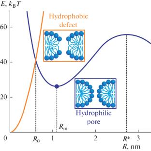
Various amphipathic antimicrobial peptides (AMPs) kill bacteria by forming through pores in plasma membranes. Previously, at least two alternative types of hypotheses about the mechanisms of AMP membrane poration were put forward. The so-called “non-local” models suggest that AMPs, when interacting with a membrane, modify its integral elastic characteristics, in particular, lateral tension, which leads to a decrease in the deformation energy during pore formation. In this case, AMP molecules can be located far from the formed pore. In “local” models, it is assumed that pores are formed in the immediate vicinity of single AMP molecules or their aggregates, while the peptides partially or completely line the edge of the pore. In both types of models, it is assumed that the process of pore formation passes via an intermediate structure, the so-called hydrophobic defect. In this work, we calculated the energy of formation of the hydrophobic defect in the membrane with adsorbed AMP molecules under the assumption of the non-local poration mechanism. It was found that AMPs actually lower the energy of the hydrophobic defect. However, this decrease in energy is insufficient to explain the experimentally observed average waiting time for membrane poration. Thus, it can be concluded that amphipathic peptides form pores in membranes predominantly by the local mechanism, directly participating in the formation of the pore edge, although nonlocal effects of AMP–membrane interaction somewhat facilitate poration of the membrane as a whole.
期刊介绍:
Biochemistry (Moscow), Supplement Series A: Membrane and Cell Biology is an international peer reviewed journal that publishes original articles on physical, chemical, and molecular mechanisms that underlie basic properties of biological membranes and mediate membrane-related cellular functions. The primary topics of the journal are membrane structure, mechanisms of membrane transport, bioenergetics and photobiology, intracellular signaling as well as membrane aspects of cell biology, immunology, and medicine. The journal is multidisciplinary and gives preference to those articles that employ a variety of experimental approaches, basically in biophysics but also in biochemistry, cytology, and molecular biology. The journal publishes articles that strive for unveiling membrane and cellular functions through innovative theoretical models and computer simulations.


 分享
分享
 求助内容:
求助内容: 应助结果提醒方式:
 扫码关注我们
