{"title":"Ultrastructure of the fertilized egg envelopes in Ancistrus cirrhosus, Loricariidae, Teleostei","authors":"Dong Heui Kim","doi":"10.1186/s42649-020-00034-7","DOIUrl":null,"url":null,"abstract":"<p>We examined the morphology of fertilized egg and ultrastructures of fertilized egg envelopes of <i>Ancistrus cirrhosus</i> belong to Loricariidae using light and electron microscopes. The fertilized eggs formed a mass on the spawning place and were yellowish, spherical, non-transparent, demersal, adhesive, and a narrow perivitelline space. But, the adhesiveness of fertilized eggs was disappeared after spawning excluding contact parts. The micropyle with funnel shape was surrounded by 15–19 furrow lines of egg envelope in a spoke-like pattern. The outer surface of egg envelope has smooth side and inner surface of egg envelope was rough with grooves. Also, the total thickness of the fertilized egg envelope was about 32.58?±?0.85?μm (<i>n</i>?=?20), and the fertilized egg envelope consisted of three layers, an outer adhesive electron-dense layer, a middle layer with low electron density and an inner electron-dense layer with grooves in counter structure from other most teleost. Collectively, these morphological characteristics and adhesive property of fertilized egg, and ultrastructures of micropyle, outer surface, and section of fertilized egg envelope are showed species specificity.</p>","PeriodicalId":470,"journal":{"name":"Applied Microscopy","volume":"50 1","pages":""},"PeriodicalIF":0.0000,"publicationDate":"2020-06-17","publicationTypes":"Journal Article","fieldsOfStudy":null,"isOpenAccess":false,"openAccessPdf":"https://sci-hub-pdf.com/10.1186/s42649-020-00034-7","citationCount":"3","resultStr":null,"platform":"Semanticscholar","paperid":null,"PeriodicalName":"Applied Microscopy","FirstCategoryId":"1085","ListUrlMain":"https://link.springer.com/article/10.1186/s42649-020-00034-7","RegionNum":0,"RegionCategory":null,"ArticlePicture":[],"TitleCN":null,"AbstractTextCN":null,"PMCID":null,"EPubDate":"","PubModel":"","JCR":"Q3","JCRName":"Immunology and Microbiology","Score":null,"Total":0}
引用次数: 3
Abstract
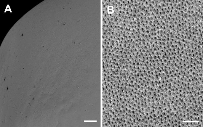
We examined the morphology of fertilized egg and ultrastructures of fertilized egg envelopes of Ancistrus cirrhosus belong to Loricariidae using light and electron microscopes. The fertilized eggs formed a mass on the spawning place and were yellowish, spherical, non-transparent, demersal, adhesive, and a narrow perivitelline space. But, the adhesiveness of fertilized eggs was disappeared after spawning excluding contact parts. The micropyle with funnel shape was surrounded by 15–19 furrow lines of egg envelope in a spoke-like pattern. The outer surface of egg envelope has smooth side and inner surface of egg envelope was rough with grooves. Also, the total thickness of the fertilized egg envelope was about 32.58?±?0.85?μm (n?=?20), and the fertilized egg envelope consisted of three layers, an outer adhesive electron-dense layer, a middle layer with low electron density and an inner electron-dense layer with grooves in counter structure from other most teleost. Collectively, these morphological characteristics and adhesive property of fertilized egg, and ultrastructures of micropyle, outer surface, and section of fertilized egg envelope are showed species specificity.
Applied MicroscopyImmunology and Microbiology-Applied Microbiology and Biotechnology
CiteScore
3.40
自引率
0.00%
发文量
10
审稿时长
10 weeks
期刊介绍:
Applied Microscopy is a peer-reviewed journal sponsored by the Korean Society of Microscopy. The journal covers all the interdisciplinary fields of technological developments in new microscopy methods and instrumentation and their applications to biological or materials science for determining structure and chemistry. ISSN: 22875123, 22874445.


 分享
分享
 求助内容:
求助内容: 应助结果提醒方式:
 扫码关注我们
