Mariana Pannerec-Varna, Philippe Ratajczak, Guilhem Bousquet, Irmine Ferreira, Christophe Leboeuf, Raphaël Boisgard, Guillaume Gapihan, Jérôme Verine, Bruno Palpant, Emmanuel Bossy, Eric Doris, Joel Poupon, Emmanuel Fort, Anne Janin
{"title":"In vivo uptake and cellular distribution of gold nanoshells in a preclinical model of xenografted human renal cancer","authors":"Mariana Pannerec-Varna, Philippe Ratajczak, Guilhem Bousquet, Irmine Ferreira, Christophe Leboeuf, Raphaël Boisgard, Guillaume Gapihan, Jérôme Verine, Bruno Palpant, Emmanuel Bossy, Eric Doris, Joel Poupon, Emmanuel Fort, Anne Janin","doi":"10.1007/s13404-013-0115-8","DOIUrl":null,"url":null,"abstract":"<p>Large-sized gold nanoparticles, promising imaging and therapeutic tools in human cancer, need long-term studies evaluating tissue bio-distribution in blood, organs and tumor. In a preclinical model of mouse xenografted with human renal cancer, we analysed the bio-distribution of a single dose (160?μg/kg) intravenously injected of poly-ethylene glycol (PEG)ylated gold nanoshells (~150?nm), in blood, normal and tumoral tissues. Using inductively coupled plasma mass spectrometry (ICP-MS), dark field and electron microscopy, we performed a sequential study of nanoshell uptake and distribution in the tumor. We also studied microscopically the organs most sensitive to efficient anticancer drugs to detect a possible long-term toxicity. Gold quantities significantly decreased in blood between early and late time points, whereas they significantly increased in liver and spleen. In addition, gold nanoshells did not induce any tissue damage, such as necrosis, inflammatory infiltrate or fibrosis in mouse liver, spleen, kidney or bone marrow after 6?months. In human renal cancer xenografts, ICP-MS showed an early decrease of gold, with 1-week stability before decrease at Day 15. Dark field microscopy showed gold particles within the vessel lumen 5 to 30?min after nanoshell injection, while 24?h later, gold particle distribution was mainly intracellular. Electron microscopy identified nanoshells within blood vessels at 5 and 30?min, within endothelial cells at 3 and 6?h and within cytoplasms of macrophages in the tumoral tissue after 24?h. In conclusion, no toxicity was observed in mice 6?months after administration of PEGylated gold nanoshells and the distribution kinetics progressed from intravascular flow at 30?min to intratumoral cells 24?h later.</p>","PeriodicalId":55086,"journal":{"name":"Gold Bulletin","volume":"46 4","pages":"257 - 265"},"PeriodicalIF":1.5000,"publicationDate":"2013-11-17","publicationTypes":"Journal Article","fieldsOfStudy":null,"isOpenAccess":false,"openAccessPdf":"https://sci-hub-pdf.com/10.1007/s13404-013-0115-8","citationCount":"16","resultStr":null,"platform":"Semanticscholar","paperid":null,"PeriodicalName":"Gold Bulletin","FirstCategoryId":"5","ListUrlMain":"https://link.springer.com/article/10.1007/s13404-013-0115-8","RegionNum":4,"RegionCategory":"工程技术","ArticlePicture":[],"TitleCN":null,"AbstractTextCN":null,"PMCID":null,"EPubDate":"","PubModel":"","JCR":"Q2","JCRName":"Chemistry","Score":null,"Total":0}
引用次数: 16
Abstract
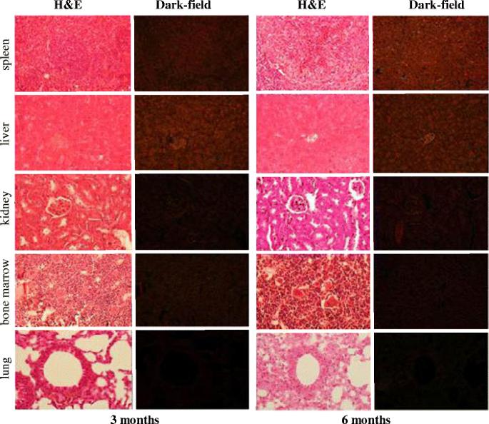
Large-sized gold nanoparticles, promising imaging and therapeutic tools in human cancer, need long-term studies evaluating tissue bio-distribution in blood, organs and tumor. In a preclinical model of mouse xenografted with human renal cancer, we analysed the bio-distribution of a single dose (160?μg/kg) intravenously injected of poly-ethylene glycol (PEG)ylated gold nanoshells (~150?nm), in blood, normal and tumoral tissues. Using inductively coupled plasma mass spectrometry (ICP-MS), dark field and electron microscopy, we performed a sequential study of nanoshell uptake and distribution in the tumor. We also studied microscopically the organs most sensitive to efficient anticancer drugs to detect a possible long-term toxicity. Gold quantities significantly decreased in blood between early and late time points, whereas they significantly increased in liver and spleen. In addition, gold nanoshells did not induce any tissue damage, such as necrosis, inflammatory infiltrate or fibrosis in mouse liver, spleen, kidney or bone marrow after 6?months. In human renal cancer xenografts, ICP-MS showed an early decrease of gold, with 1-week stability before decrease at Day 15. Dark field microscopy showed gold particles within the vessel lumen 5 to 30?min after nanoshell injection, while 24?h later, gold particle distribution was mainly intracellular. Electron microscopy identified nanoshells within blood vessels at 5 and 30?min, within endothelial cells at 3 and 6?h and within cytoplasms of macrophages in the tumoral tissue after 24?h. In conclusion, no toxicity was observed in mice 6?months after administration of PEGylated gold nanoshells and the distribution kinetics progressed from intravascular flow at 30?min to intratumoral cells 24?h later.
期刊介绍:
Gold Bulletin is the premier international peer reviewed journal on the latest science, technology and applications of gold. It includes papers on the latest research advances, state-of-the-art reviews, conference reports, book reviews and highlights of patents and scientific literature. Gold Bulletin does not publish manuscripts covering the snthesis of Gold nanoparticles in the presence of plant extracts or other nature-derived extracts. Gold Bulletin has been published over 40 years as a multidisciplinary journal read by chemists, physicists, engineers, metallurgists, materials scientists, biotechnologists, surface scientists, and nanotechnologists amongst others, both within industry and academia. Gold Bulletin is published in Association with the World Gold Council.


 分享
分享
 求助内容:
求助内容: 应助结果提醒方式:
 扫码关注我们
