Byung Soo Chang, Eun-Kyung Choi, Hyun-Wook Kim, Dong Heui Kim
{"title":"Ultrastructure of the fertilized egg envelopes in Hemigrammus erythrozonus, Characidae, Teleostei","authors":"Byung Soo Chang, Eun-Kyung Choi, Hyun-Wook Kim, Dong Heui Kim","doi":"10.1186/s42649-019-0010-8","DOIUrl":null,"url":null,"abstract":"<p>We examined the morphology and ultrastructures of fertilized egg envelopes of glowlight tetra (<i>Hemigrammus erythrozonus</i>) belong to Characidae using light and electron microscopes.</p><p>The fertilized eggs were spherical, transparent, demersal, adhesive, and have no oil droplet. The perivitelline space was well-developed and the micropyle was surrounded by 15–20 uplifted lines of egg envelope in a spoke like pattern. The outer surface of egg envelope was rough side with grooves. Also, the total thickness of the fertilized egg envelope was about 2.1–2.3?μm, and the fertilized egg envelope consisted of two layers, an outer adhesive electron-dense layer with grooves and three feather-like lamellae layers. Collectively, these morphological characteristics of fertilized egg and micropyle with spoke-like structure showed family Characidae specificity, and ultrastructures of outer surface and section of fertilized egg envelope showed species specificity.</p>","PeriodicalId":470,"journal":{"name":"Applied Microscopy","volume":"49 1","pages":""},"PeriodicalIF":0.0000,"publicationDate":"2019-08-20","publicationTypes":"Journal Article","fieldsOfStudy":null,"isOpenAccess":false,"openAccessPdf":"https://sci-hub-pdf.com/10.1186/s42649-019-0010-8","citationCount":"3","resultStr":null,"platform":"Semanticscholar","paperid":null,"PeriodicalName":"Applied Microscopy","FirstCategoryId":"1085","ListUrlMain":"https://link.springer.com/article/10.1186/s42649-019-0010-8","RegionNum":0,"RegionCategory":null,"ArticlePicture":[],"TitleCN":null,"AbstractTextCN":null,"PMCID":null,"EPubDate":"","PubModel":"","JCR":"Q3","JCRName":"Immunology and Microbiology","Score":null,"Total":0}
引用次数: 3
Abstract
We examined the morphology and ultrastructures of fertilized egg envelopes of glowlight tetra (Hemigrammus erythrozonus) belong to Characidae using light and electron microscopes.
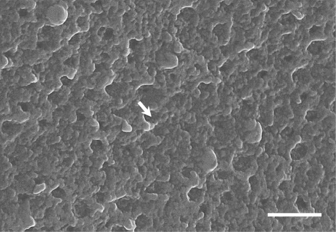
The fertilized eggs were spherical, transparent, demersal, adhesive, and have no oil droplet. The perivitelline space was well-developed and the micropyle was surrounded by 15–20 uplifted lines of egg envelope in a spoke like pattern. The outer surface of egg envelope was rough side with grooves. Also, the total thickness of the fertilized egg envelope was about 2.1–2.3?μm, and the fertilized egg envelope consisted of two layers, an outer adhesive electron-dense layer with grooves and three feather-like lamellae layers. Collectively, these morphological characteristics of fertilized egg and micropyle with spoke-like structure showed family Characidae specificity, and ultrastructures of outer surface and section of fertilized egg envelope showed species specificity.
Applied MicroscopyImmunology and Microbiology-Applied Microbiology and Biotechnology
CiteScore
3.40
自引率
0.00%
发文量
10
审稿时长
10 weeks
期刊介绍:
Applied Microscopy is a peer-reviewed journal sponsored by the Korean Society of Microscopy. The journal covers all the interdisciplinary fields of technological developments in new microscopy methods and instrumentation and their applications to biological or materials science for determining structure and chemistry. ISSN: 22875123, 22874445.


 分享
分享
 求助内容:
求助内容: 应助结果提醒方式:
 扫码关注我们
