Feng Wang, Trond R. Henninen, Debora Keller, Rolf Erni
{"title":"Noise2Atom: unsupervised denoising for scanning transmission electron microscopy images","authors":"Feng Wang, Trond R. Henninen, Debora Keller, Rolf Erni","doi":"10.1186/s42649-020-00041-8","DOIUrl":null,"url":null,"abstract":"<p>We propose an effective deep learning model to denoise scanning transmission electron microscopy (STEM) image series, named Noise2Atom, to map images from a source domain <span>\\(\\mathcal {S}\\)</span> to a target domain <span>\\(\\mathcal {C}\\)</span>, where <span>\\(\\mathcal {S}\\)</span> is for our noisy experimental dataset, and <span>\\(\\mathcal {C}\\)</span> is for the desired clear atomic images. Noise2Atom uses two external networks to apply additional constraints from the domain knowledge. This model requires no signal prior, no noise model estimation, and no paired training images. The only assumption is that the inputs are acquired with identical experimental configurations. To evaluate the restoration performance of our model, as it is impossible to obtain ground truth for our experimental dataset, we propose consecutive structural similarity (CSS) for image quality assessment, based on the fact that the structures remain much the same as the previous frame(s) within small scan intervals. We demonstrate the superiority of our model by providing evaluation in terms of CSS and visual quality on different experimental datasets.</p>","PeriodicalId":470,"journal":{"name":"Applied Microscopy","volume":"50 1","pages":""},"PeriodicalIF":0.0000,"publicationDate":"2020-10-20","publicationTypes":"Journal Article","fieldsOfStudy":null,"isOpenAccess":false,"openAccessPdf":"https://sci-hub-pdf.com/10.1186/s42649-020-00041-8","citationCount":"28","resultStr":null,"platform":"Semanticscholar","paperid":null,"PeriodicalName":"Applied Microscopy","FirstCategoryId":"1085","ListUrlMain":"https://link.springer.com/article/10.1186/s42649-020-00041-8","RegionNum":0,"RegionCategory":null,"ArticlePicture":[],"TitleCN":null,"AbstractTextCN":null,"PMCID":null,"EPubDate":"","PubModel":"","JCR":"Q3","JCRName":"Immunology and Microbiology","Score":null,"Total":0}
引用次数: 28
Abstract
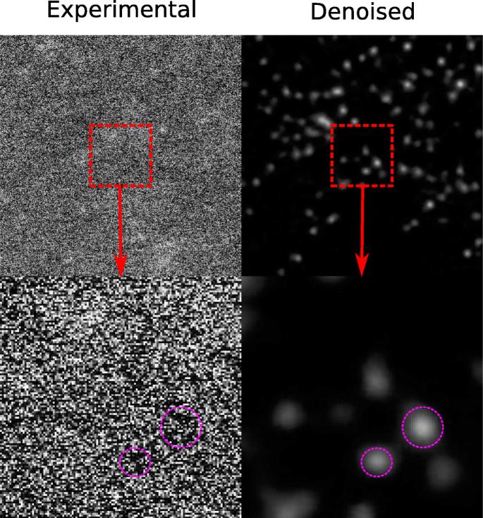
We propose an effective deep learning model to denoise scanning transmission electron microscopy (STEM) image series, named Noise2Atom, to map images from a source domain \(\mathcal {S}\) to a target domain \(\mathcal {C}\), where \(\mathcal {S}\) is for our noisy experimental dataset, and \(\mathcal {C}\) is for the desired clear atomic images. Noise2Atom uses two external networks to apply additional constraints from the domain knowledge. This model requires no signal prior, no noise model estimation, and no paired training images. The only assumption is that the inputs are acquired with identical experimental configurations. To evaluate the restoration performance of our model, as it is impossible to obtain ground truth for our experimental dataset, we propose consecutive structural similarity (CSS) for image quality assessment, based on the fact that the structures remain much the same as the previous frame(s) within small scan intervals. We demonstrate the superiority of our model by providing evaluation in terms of CSS and visual quality on different experimental datasets.
Applied MicroscopyImmunology and Microbiology-Applied Microbiology and Biotechnology
CiteScore
3.40
自引率
0.00%
发文量
10
审稿时长
10 weeks
期刊介绍:
Applied Microscopy is a peer-reviewed journal sponsored by the Korean Society of Microscopy. The journal covers all the interdisciplinary fields of technological developments in new microscopy methods and instrumentation and their applications to biological or materials science for determining structure and chemistry. ISSN: 22875123, 22874445.


 分享
分享
 求助内容:
求助内容: 应助结果提醒方式:
 扫码关注我们
