Ji Hea Bang, Jin Hyong Kang, Seung Han Kim, Jong Jae Park, Beom Jae Lee, Moon Kyung Joo, Tae Sung Jeon
{"title":"Primary Esophageal Mucosa-associated Lymphoid Tissue Lymphoma: A Rare Case Report and Review of Other Published Data.","authors":"Ji Hea Bang, Jin Hyong Kang, Seung Han Kim, Jong Jae Park, Beom Jae Lee, Moon Kyung Joo, Tae Sung Jeon","doi":"10.7704/kjhugr.2023.0026","DOIUrl":null,"url":null,"abstract":"<p><p>The gastrointestinal tract is the most common extranodal site for lymphomas, and mucosa-associated lymphoid tissue lymphoma is the second most common histological lymphoma subtype. However, primary esophageal mucosa-associated lymphoid tissue lymphomas are extremely rare. Few such cases are documented, and the reports demonstrate inconsistent diagnostic and therapeutic strategies. Herein, a 54-year-old man was referred to our hospital for treatment of dysphagia. Esophagogastroduodenoscopy revealed a large, horseshoe-shaped subepithelial mass in the upper esophagus. Endoscopic ultrasonography and computed tomography revealed that the mass was well-demarcated and confined to the muscularis mucosa, with no abnormalities in other organs or lymph nodes. The mass was presumptively diagnosed as benign, and the patient underwent endoscopic mucosal dissection for pathological confirmation and symptom relief. Pathological examination of the dissection specimen revealed that it was a primary esophageal mucosa-associated lymphoid tissue lymphoma. As the patient had an elevated immunoglobulin G level and <i>Helicobacter pylori</i> infection, we administered adjuvant eradication therapy. The patient remains under surveillance and is free of lymphoma recurrence 36 months postoperatively. This case report demonstrates that endoscopic resection and <i>H. pylori</i> eradication are effective treatment strategies for early-stage esophageal mucosa-associated lymphoid tissue lymphoma.</p>","PeriodicalId":22895,"journal":{"name":"The Korean Journal of Helicobacter and Upper Gastrointestinal Research","volume":" ","pages":"207-213"},"PeriodicalIF":0.0000,"publicationDate":"2023-09-01","publicationTypes":"Journal Article","fieldsOfStudy":null,"isOpenAccess":false,"openAccessPdf":"https://www.ncbi.nlm.nih.gov/pmc/articles/PMC11967450/pdf/","citationCount":"0","resultStr":null,"platform":"Semanticscholar","paperid":null,"PeriodicalName":"The Korean Journal of Helicobacter and Upper Gastrointestinal Research","FirstCategoryId":"1085","ListUrlMain":"https://doi.org/10.7704/kjhugr.2023.0026","RegionNum":0,"RegionCategory":null,"ArticlePicture":[],"TitleCN":null,"AbstractTextCN":null,"PMCID":null,"EPubDate":"2023/8/30 0:00:00","PubModel":"Epub","JCR":"","JCRName":"","Score":null,"Total":0}
引用次数: 0
Abstract
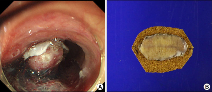
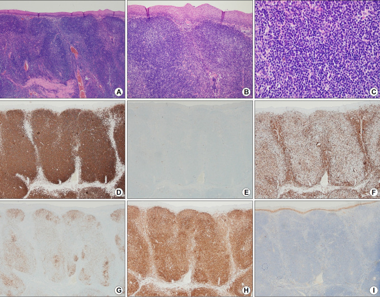
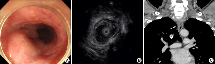
The gastrointestinal tract is the most common extranodal site for lymphomas, and mucosa-associated lymphoid tissue lymphoma is the second most common histological lymphoma subtype. However, primary esophageal mucosa-associated lymphoid tissue lymphomas are extremely rare. Few such cases are documented, and the reports demonstrate inconsistent diagnostic and therapeutic strategies. Herein, a 54-year-old man was referred to our hospital for treatment of dysphagia. Esophagogastroduodenoscopy revealed a large, horseshoe-shaped subepithelial mass in the upper esophagus. Endoscopic ultrasonography and computed tomography revealed that the mass was well-demarcated and confined to the muscularis mucosa, with no abnormalities in other organs or lymph nodes. The mass was presumptively diagnosed as benign, and the patient underwent endoscopic mucosal dissection for pathological confirmation and symptom relief. Pathological examination of the dissection specimen revealed that it was a primary esophageal mucosa-associated lymphoid tissue lymphoma. As the patient had an elevated immunoglobulin G level and Helicobacter pylori infection, we administered adjuvant eradication therapy. The patient remains under surveillance and is free of lymphoma recurrence 36 months postoperatively. This case report demonstrates that endoscopic resection and H. pylori eradication are effective treatment strategies for early-stage esophageal mucosa-associated lymphoid tissue lymphoma.


 分享
分享

 求助内容:
求助内容: 应助结果提醒方式:
 扫码关注我们
