{"title":"Microscopic research on the olfactory organ of the Far Eastern brook lamprey Lethenteron reissneri (Pisces, Petromyzontidae)","authors":"Hyun-Tae Kim, Jong-Young Park","doi":"10.1186/s42649-020-00038-3","DOIUrl":null,"url":null,"abstract":"<p>The olfactory anatomy and histology of <i>Lethenteron reissneri</i> were researched using a stereo microscope, a light microscope, and a scanning electron microscope. As in other lampreys, it shows same characters as follows: i) a single olfactory organ, ii) a single tubular nostril, iii) a single olfactory chamber with gourd-like form, iv) a nasal valve, v) a nasopharyngeal pouch, vi) a sensory epithelium (SE) of continuous distribution, vii) a supporting cells with numerous long cilia, viii) an accessory olfactory organ. However, the description of a pseudostratified columnar layer in the SE and Non SE is a first record, not reported in sea lamprey <i>Petromyzon marinus</i>. In particular, both 19 to 20 lamellae in number and olfactory receptor neuron’s quarter ciliary length of the knob diameter differ from those of <i>P. marinus</i>. From these results, it might be considered that the olfactory organ of <i>L. reissneri</i> shows well adaptive structure of a primitive fish to slow flowing water with gravel, pebbles, and sand and a hiding habit into sand bottom at daytime. The lamellar number and neuron’s ciliary length may be a meaningful taxonomic character for the class Petromyzonida.</p>","PeriodicalId":470,"journal":{"name":"Applied Microscopy","volume":"50 1","pages":""},"PeriodicalIF":0.0000,"publicationDate":"2020-09-15","publicationTypes":"Journal Article","fieldsOfStudy":null,"isOpenAccess":false,"openAccessPdf":"https://sci-hub-pdf.com/10.1186/s42649-020-00038-3","citationCount":"1","resultStr":null,"platform":"Semanticscholar","paperid":null,"PeriodicalName":"Applied Microscopy","FirstCategoryId":"1085","ListUrlMain":"https://link.springer.com/article/10.1186/s42649-020-00038-3","RegionNum":0,"RegionCategory":null,"ArticlePicture":[],"TitleCN":null,"AbstractTextCN":null,"PMCID":null,"EPubDate":"","PubModel":"","JCR":"Q3","JCRName":"Immunology and Microbiology","Score":null,"Total":0}
引用次数: 1
Abstract
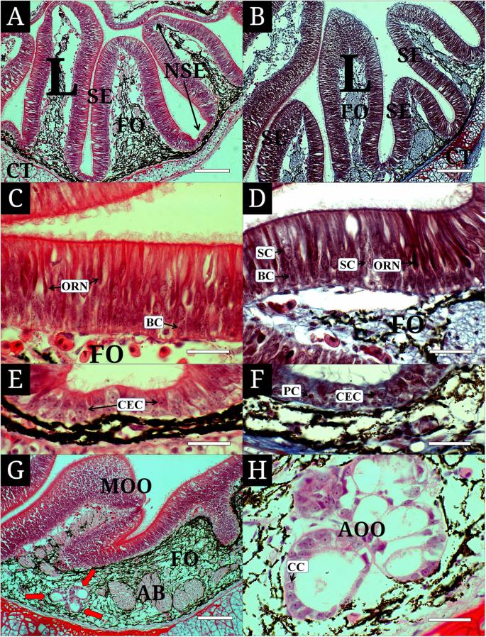
The olfactory anatomy and histology of Lethenteron reissneri were researched using a stereo microscope, a light microscope, and a scanning electron microscope. As in other lampreys, it shows same characters as follows: i) a single olfactory organ, ii) a single tubular nostril, iii) a single olfactory chamber with gourd-like form, iv) a nasal valve, v) a nasopharyngeal pouch, vi) a sensory epithelium (SE) of continuous distribution, vii) a supporting cells with numerous long cilia, viii) an accessory olfactory organ. However, the description of a pseudostratified columnar layer in the SE and Non SE is a first record, not reported in sea lamprey Petromyzon marinus. In particular, both 19 to 20 lamellae in number and olfactory receptor neuron’s quarter ciliary length of the knob diameter differ from those of P. marinus. From these results, it might be considered that the olfactory organ of L. reissneri shows well adaptive structure of a primitive fish to slow flowing water with gravel, pebbles, and sand and a hiding habit into sand bottom at daytime. The lamellar number and neuron’s ciliary length may be a meaningful taxonomic character for the class Petromyzonida.
Applied MicroscopyImmunology and Microbiology-Applied Microbiology and Biotechnology
CiteScore
3.40
自引率
0.00%
发文量
10
审稿时长
10 weeks
期刊介绍:
Applied Microscopy is a peer-reviewed journal sponsored by the Korean Society of Microscopy. The journal covers all the interdisciplinary fields of technological developments in new microscopy methods and instrumentation and their applications to biological or materials science for determining structure and chemistry. ISSN: 22875123, 22874445.


 分享
分享
 求助内容:
求助内容: 应助结果提醒方式:
 扫码关注我们
