Crissy-Ann Harrylal, Antoinette V. Lensink, Sunil K. Gupta, Tom A. Aire
{"title":"The ultrastructural features of the infundibulum of the green iguana, Iguana iguana","authors":"Crissy-Ann Harrylal, Antoinette V. Lensink, Sunil K. Gupta, Tom A. Aire","doi":"10.1002/jmor.21644","DOIUrl":null,"url":null,"abstract":"<div>\n \n <p>The purpose of this study is to describe, in detail, the ultrastructure of the infundibulum of the sexually mature and active female green iguana, <i>Iguana iguana</i>. The infundibulum of five iguanas was remarkably distinct from the uterus, and was also clearly demarcated into cranial (expanded v-shaped) and caudal (tubular) divisions. Tissue samples obtained from five portions (three from the cranial division and two from the caudal division) of the infundibulum were processed conventionally for light and electron microscopy. The epithelial lining of the most anterior, middle, and posterior, parts of the cranial division displayed nonciliated cells predominantly, and occasionally ciliated cells. The numerous secretory granules in nonciliated type 1 cell found in the fimbrial aspect of the infundibulum were homogenous and deeply electron-dense, but those in the other two regions were variants of this cell type because they contained variably electron-dense secretory granules. Two main types of nonciliated cells (type 2 and its variant, type 3, as well as type 4) occurred in the epithelial lining of the caudal division of the infundibulum, but they, clearly, showed no dense secretory granules. Whereas the nonciliated type 2 cell and its variant (type 3 cell) contained large glycogen deposits, the type 4 cell lacked these deposits but its apical part contained large lipid-like droplets and, remarkably, blebbed into the duct lumen. The nonciliated cells lining the mucosal tubular glands contained highly electron-dense secretory granules, which were similar to those found in the nonciliated type 1 cell in the epithelial lining of the fimbrial part of the cranial division of the infundibulum.</p></div>","PeriodicalId":16528,"journal":{"name":"Journal of Morphology","volume":"284 11","pages":""},"PeriodicalIF":1.4000,"publicationDate":"2023-10-08","publicationTypes":"Journal Article","fieldsOfStudy":null,"isOpenAccess":false,"openAccessPdf":"","citationCount":"0","resultStr":null,"platform":"Semanticscholar","paperid":null,"PeriodicalName":"Journal of Morphology","FirstCategoryId":"3","ListUrlMain":"https://onlinelibrary.wiley.com/doi/10.1002/jmor.21644","RegionNum":4,"RegionCategory":"医学","ArticlePicture":[],"TitleCN":null,"AbstractTextCN":null,"PMCID":null,"EPubDate":"","PubModel":"","JCR":"Q2","JCRName":"ANATOMY & MORPHOLOGY","Score":null,"Total":0}
引用次数: 0
Abstract
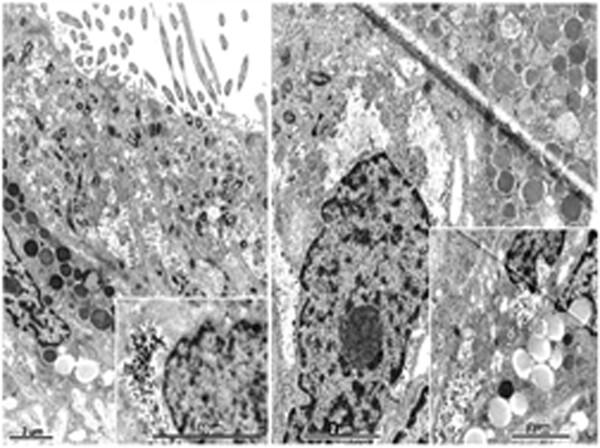
The purpose of this study is to describe, in detail, the ultrastructure of the infundibulum of the sexually mature and active female green iguana, Iguana iguana. The infundibulum of five iguanas was remarkably distinct from the uterus, and was also clearly demarcated into cranial (expanded v-shaped) and caudal (tubular) divisions. Tissue samples obtained from five portions (three from the cranial division and two from the caudal division) of the infundibulum were processed conventionally for light and electron microscopy. The epithelial lining of the most anterior, middle, and posterior, parts of the cranial division displayed nonciliated cells predominantly, and occasionally ciliated cells. The numerous secretory granules in nonciliated type 1 cell found in the fimbrial aspect of the infundibulum were homogenous and deeply electron-dense, but those in the other two regions were variants of this cell type because they contained variably electron-dense secretory granules. Two main types of nonciliated cells (type 2 and its variant, type 3, as well as type 4) occurred in the epithelial lining of the caudal division of the infundibulum, but they, clearly, showed no dense secretory granules. Whereas the nonciliated type 2 cell and its variant (type 3 cell) contained large glycogen deposits, the type 4 cell lacked these deposits but its apical part contained large lipid-like droplets and, remarkably, blebbed into the duct lumen. The nonciliated cells lining the mucosal tubular glands contained highly electron-dense secretory granules, which were similar to those found in the nonciliated type 1 cell in the epithelial lining of the fimbrial part of the cranial division of the infundibulum.
期刊介绍:
The Journal of Morphology welcomes articles of original research in cytology, protozoology, embryology, and general morphology. Articles generally should not exceed 35 printed pages. Preliminary notices or articles of a purely descriptive morphological or taxonomic nature are not included. No paper which has already been published will be accepted, nor will simultaneous publications elsewhere be allowed.
The Journal of Morphology publishes research in functional, comparative, evolutionary and developmental morphology from vertebrates and invertebrates. Human and veterinary anatomy or paleontology are considered when an explicit connection to neontological animal morphology is presented, and the paper contains relevant information for the community of animal morphologists. Based on our long tradition, we continue to seek publishing the best papers in animal morphology.


 分享
分享
 求助内容:
求助内容: 应助结果提醒方式:
 扫码关注我们
