{"title":"Atypical azygos continuation of the caudal vena cava in a dog","authors":"A. Masuyama, A. Sato, M. Murakami","doi":"10.1111/jsap.13686","DOIUrl":null,"url":null,"abstract":"<p>A whole-body CT scan and CT myelography were performed on a 13-year-old male neutered Pug with gait abnormality and a history of cough that had been medically managed. The CT scan revealed multiple sites of intervertebral disc disease and incidentally revealed an abnormally distended azygos vein with absent pre-hepatic caudal vena cava (CVC). The pre-renal segment of the CVC was duplicated with both common iliac veins joining to each duplicated CVC, and then cranially connected to the azygos vein (cavo-right-azygos shunt) (Fig 1A, B). Both renal veins connected and then joining to a cranially coursing tortuous shunting vessel connecting to the azygos vein in the thoracic cavity (Fig 1A, B, D). Thus, the renal veins were not directly connected to the pre-renal segment of CVC in the abdomen. The hepatic and post-hepatic segments of the CVC were present as normal. Segmental aplasia of the CVC with azygos continuation is a congenital anomaly characterised by the CVC uniting the azygos vein with partial absence of the CVC. In this case, concurrent partial duplication of the CVC was present. The duplicated pre-renal segment of CVC course dorsal to the aorta and cranially continued to the azygos vein. To the best of the authors' knowledge, the atypical azygos continuation of the CVC with concurrent segmental aplasia of the pre-hepatic CVC has not been reported. Although concomitant portosystemic shunting is common in azygos continuation of the CVC, there was no evidence of portosystemic shunting in the present case. This dog had no clinical signs related to this anomaly; therefore, it was considered incidental, similar to most cases of azygos continuation of the CVC.</p><p>The work described in this manuscript involved the use of a non-experimental (owned) animal. Established internationally recognised high standards (‘best practice’) of veterinary clinical care for the individual patient were always followed. Ethical approval from a committee was therefore not specifically required for publication in Images in Small Animal Practice in Journal of Small Animal Practice. Although not required, ethical approval was still obtained.</p>","PeriodicalId":17062,"journal":{"name":"Journal of Small Animal Practice","volume":"65 2","pages":"150"},"PeriodicalIF":1.9000,"publicationDate":"2023-10-22","publicationTypes":"Journal Article","fieldsOfStudy":null,"isOpenAccess":false,"openAccessPdf":"https://onlinelibrary.wiley.com/doi/epdf/10.1111/jsap.13686","citationCount":"0","resultStr":null,"platform":"Semanticscholar","paperid":null,"PeriodicalName":"Journal of Small Animal Practice","FirstCategoryId":"97","ListUrlMain":"https://onlinelibrary.wiley.com/doi/10.1111/jsap.13686","RegionNum":2,"RegionCategory":"农林科学","ArticlePicture":[],"TitleCN":null,"AbstractTextCN":null,"PMCID":null,"EPubDate":"","PubModel":"","JCR":"Q2","JCRName":"VETERINARY SCIENCES","Score":null,"Total":0}
引用次数: 0
Abstract
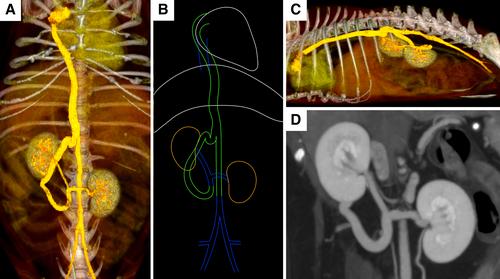
A whole-body CT scan and CT myelography were performed on a 13-year-old male neutered Pug with gait abnormality and a history of cough that had been medically managed. The CT scan revealed multiple sites of intervertebral disc disease and incidentally revealed an abnormally distended azygos vein with absent pre-hepatic caudal vena cava (CVC). The pre-renal segment of the CVC was duplicated with both common iliac veins joining to each duplicated CVC, and then cranially connected to the azygos vein (cavo-right-azygos shunt) (Fig 1A, B). Both renal veins connected and then joining to a cranially coursing tortuous shunting vessel connecting to the azygos vein in the thoracic cavity (Fig 1A, B, D). Thus, the renal veins were not directly connected to the pre-renal segment of CVC in the abdomen. The hepatic and post-hepatic segments of the CVC were present as normal. Segmental aplasia of the CVC with azygos continuation is a congenital anomaly characterised by the CVC uniting the azygos vein with partial absence of the CVC. In this case, concurrent partial duplication of the CVC was present. The duplicated pre-renal segment of CVC course dorsal to the aorta and cranially continued to the azygos vein. To the best of the authors' knowledge, the atypical azygos continuation of the CVC with concurrent segmental aplasia of the pre-hepatic CVC has not been reported. Although concomitant portosystemic shunting is common in azygos continuation of the CVC, there was no evidence of portosystemic shunting in the present case. This dog had no clinical signs related to this anomaly; therefore, it was considered incidental, similar to most cases of azygos continuation of the CVC.
The work described in this manuscript involved the use of a non-experimental (owned) animal. Established internationally recognised high standards (‘best practice’) of veterinary clinical care for the individual patient were always followed. Ethical approval from a committee was therefore not specifically required for publication in Images in Small Animal Practice in Journal of Small Animal Practice. Although not required, ethical approval was still obtained.
期刊介绍:
Journal of Small Animal Practice (JSAP) is a monthly peer-reviewed publication integrating clinical research papers and case reports from international sources, covering all aspects of medicine and surgery relating to dogs, cats and other small animals. These papers facilitate the dissemination and implementation of new ideas and techniques relating to clinical veterinary practice, with the ultimate aim of promoting best practice. JSAP publishes high quality original articles, as well as other scientific and educational information. New developments are placed in perspective, encompassing new concepts and peer commentary. The target audience is veterinarians primarily engaged in the practise of small animal medicine and surgery.
In addition to original articles, JSAP will publish invited editorials (relating to a manuscript in the same issue or a topic of current interest), review articles, which provide in-depth discussion of important clinical issues, and other scientific and educational information from around the world.
The final decision on publication of a manuscript rests with the Editorial Board and ultimately with the Editor. All papers, regardless of type, represent the opinion of the authors and not necessarily that of the Editor, the Association or the Publisher.
The Journal of Small Animal Practice is published on behalf of the British Small Animal Veterinary Association and is also the official scientific journal of the World Small Animal Veterinary Association


 分享
分享
 求助内容:
求助内容: 应助结果提醒方式:
 扫码关注我们
