Osman Kanatsız, Filiz Özülker, Tamer Aydın, Tamer Özülker
{"title":"Isolated Castrate-resistant Prostate Cancer Metastasis to Both Adrenal Glands Detected on <sup>68</sup>Ga PSMA PET/CT.","authors":"Osman Kanatsız, Filiz Özülker, Tamer Aydın, Tamer Özülker","doi":"10.4274/mirt.galenos.2023.03780","DOIUrl":null,"url":null,"abstract":"<p><p>A 61-year-old male patient, who had undergone radical prostatectomy, underwent <sup>68</sup>Ga labeled prostate-specific membrane antigen (PSMA) positron emission tomography/computerized tomography (PET/CT) for evaluation of suspected biochemical recurrence of prostate cancer (PCa). PET/CT scan showed increased <sup>68</sup>Ga PSMA expressions in hypodense mass lesions in both adrenal gland localizations. An adrenal gland tru-cut biopsy was performed for the right side, which showed poor-differentiated carcinoma metastases associated with the patient's high-grade PCa. As far as we could determine based on an extensive literature search, this is the second case in which isolated adrenal metastasis was detected by <sup>68</sup>Ga PSMA PET/CT study in a patient with PCa.</p>","PeriodicalId":44681,"journal":{"name":"Molecular Imaging and Radionuclide Therapy","volume":"32 3","pages":"244-246"},"PeriodicalIF":1.1000,"publicationDate":"2023-10-20","publicationTypes":"Journal Article","fieldsOfStudy":null,"isOpenAccess":false,"openAccessPdf":"https://ftp.ncbi.nlm.nih.gov/pub/pmc/oa_pdf/af/b4/MIRT-32-244.PMC10600555.pdf","citationCount":"0","resultStr":null,"platform":"Semanticscholar","paperid":null,"PeriodicalName":"Molecular Imaging and Radionuclide Therapy","FirstCategoryId":"1085","ListUrlMain":"https://doi.org/10.4274/mirt.galenos.2023.03780","RegionNum":0,"RegionCategory":null,"ArticlePicture":[],"TitleCN":null,"AbstractTextCN":null,"PMCID":null,"EPubDate":"","PubModel":"","JCR":"Q4","JCRName":"RADIOLOGY, NUCLEAR MEDICINE & MEDICAL IMAGING","Score":null,"Total":0}
引用次数: 0
Abstract
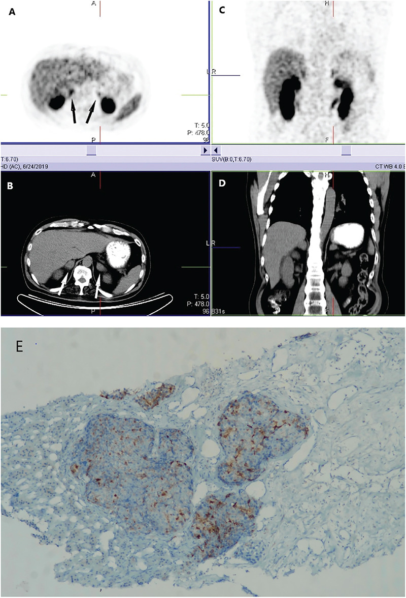
A 61-year-old male patient, who had undergone radical prostatectomy, underwent 68Ga labeled prostate-specific membrane antigen (PSMA) positron emission tomography/computerized tomography (PET/CT) for evaluation of suspected biochemical recurrence of prostate cancer (PCa). PET/CT scan showed increased 68Ga PSMA expressions in hypodense mass lesions in both adrenal gland localizations. An adrenal gland tru-cut biopsy was performed for the right side, which showed poor-differentiated carcinoma metastases associated with the patient's high-grade PCa. As far as we could determine based on an extensive literature search, this is the second case in which isolated adrenal metastasis was detected by 68Ga PSMA PET/CT study in a patient with PCa.
期刊介绍:
Molecular Imaging and Radionuclide Therapy (Mol Imaging Radionucl Ther, MIRT) is publishes original research articles, invited reviews, editorials, short communications, letters, consensus statements, guidelines and case reports with a literature review on the topic, in the field of molecular imaging, multimodality imaging, nuclear medicine, radionuclide therapy, radiopharmacy, medical physics, dosimetry and radiobiology.


 分享
分享
 求助内容:
求助内容: 应助结果提醒方式:
 扫码关注我们
