Refia Yükseltürk, Aslıhan Yıldırım, Nedim C M Gülaldı
{"title":"PET/CT Imaging of Inflammatory Myofibroblastic Tumor of the Thigh.","authors":"Refia Yükseltürk, Aslıhan Yıldırım, Nedim C M Gülaldı","doi":"10.4274/mirt.galenos.2023.55798","DOIUrl":null,"url":null,"abstract":"<p><p>A 13-year-old male patient presented with right leg pain and walking difficulty. Contrast-enhanced magnetic resonance imaging showed a softtissue lesion between muscle groups in the anterior half of the right thigh. The excisional biopsy result ended in an inflammatory myofibroblastic tumor (IMT). The <sup>18</sup>F-fluorodeoxyglucose positron emission tomography/computed tomography (PET/CT) scan showed hypermetabolism in the multifocal soft tissue lesion and also confirmed that no other distant foci were present. A three-phase Tc-99m-methylene diphosphonate study of the region showed heterogeneously increased vascularity within the region. We described an unusual case of IMT in a pediatric patient and the importance of PET/CT scanning to delineate the lesion.</p>","PeriodicalId":44681,"journal":{"name":"Molecular Imaging and Radionuclide Therapy","volume":"32 3","pages":"239-243"},"PeriodicalIF":1.1000,"publicationDate":"2023-10-20","publicationTypes":"Journal Article","fieldsOfStudy":null,"isOpenAccess":false,"openAccessPdf":"https://ftp.ncbi.nlm.nih.gov/pub/pmc/oa_pdf/59/10/MIRT-32-239.PMC10600557.pdf","citationCount":"0","resultStr":null,"platform":"Semanticscholar","paperid":null,"PeriodicalName":"Molecular Imaging and Radionuclide Therapy","FirstCategoryId":"1085","ListUrlMain":"https://doi.org/10.4274/mirt.galenos.2023.55798","RegionNum":0,"RegionCategory":null,"ArticlePicture":[],"TitleCN":null,"AbstractTextCN":null,"PMCID":null,"EPubDate":"","PubModel":"","JCR":"Q4","JCRName":"RADIOLOGY, NUCLEAR MEDICINE & MEDICAL IMAGING","Score":null,"Total":0}
引用次数: 0
Abstract
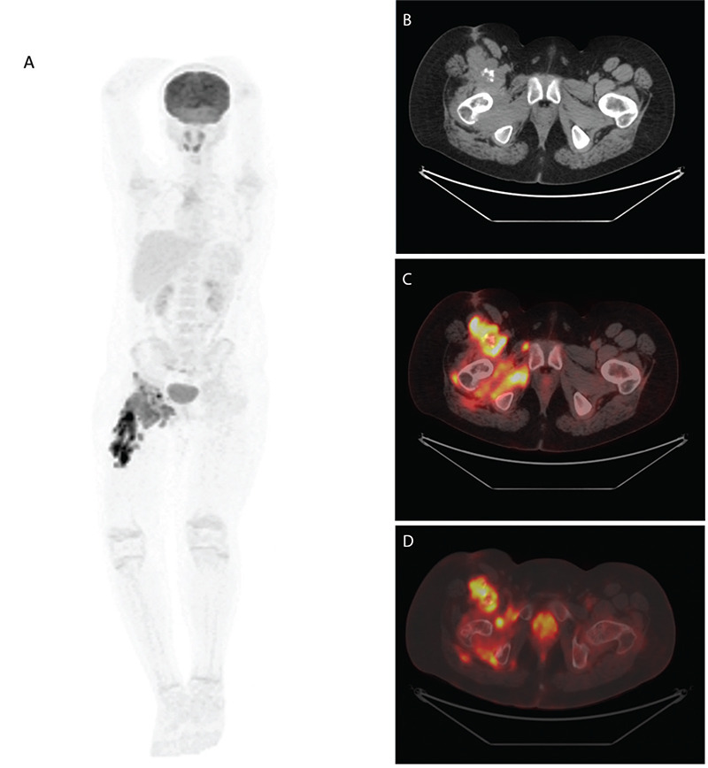
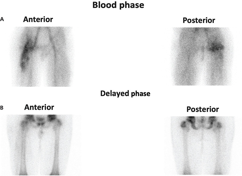
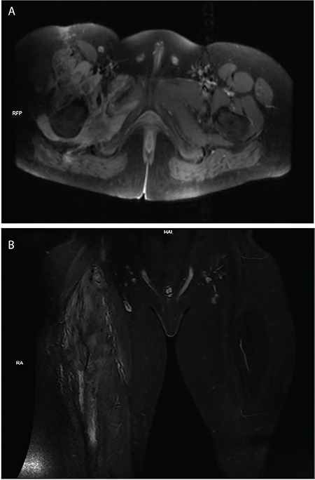
A 13-year-old male patient presented with right leg pain and walking difficulty. Contrast-enhanced magnetic resonance imaging showed a softtissue lesion between muscle groups in the anterior half of the right thigh. The excisional biopsy result ended in an inflammatory myofibroblastic tumor (IMT). The 18F-fluorodeoxyglucose positron emission tomography/computed tomography (PET/CT) scan showed hypermetabolism in the multifocal soft tissue lesion and also confirmed that no other distant foci were present. A three-phase Tc-99m-methylene diphosphonate study of the region showed heterogeneously increased vascularity within the region. We described an unusual case of IMT in a pediatric patient and the importance of PET/CT scanning to delineate the lesion.
期刊介绍:
Molecular Imaging and Radionuclide Therapy (Mol Imaging Radionucl Ther, MIRT) is publishes original research articles, invited reviews, editorials, short communications, letters, consensus statements, guidelines and case reports with a literature review on the topic, in the field of molecular imaging, multimodality imaging, nuclear medicine, radionuclide therapy, radiopharmacy, medical physics, dosimetry and radiobiology.


 分享
分享
 求助内容:
求助内容: 应助结果提醒方式:
 扫码关注我们
