求助PDF
{"title":"Validation of the utilisation of automatic placement of anatomical and sliding landmarks on three-dimensional models for shape analysis of human pelves","authors":"TM Mbonani , AC Hagg , EN L'Abbé , AC Oettlé , AF Ridel","doi":"10.1016/j.fri.2023.200542","DOIUrl":null,"url":null,"abstract":"<div><p>Estimating sex from unknown human skeletal remains is an important component in forensic anthropology. Currently, both morphological and morphometric methods are used for sex estimation. These methods employ landmarks to make morphological comparisons between and within groups. Manual landmarking has been regarded as time-consuming and subjective. To decrease observer subjectivity and reduce measurement errors, an automated three-dimensional (3D) method was developed. This study aimed to validate the utilisation of the automatic placement of anatomical and sliding landmarks on 3D pelvis models for shape analysis using Computed Tomography (CT) scans.</p><p>Computed Tomography scans of adult South Africans were obtained from Steve Biko Academic Hospital, Pretoria, South Africa. In this study, automatic landmarking was validated on 130 3D reconstructions of the adult human pelvis. Eighteen anatomical and 260 sliding landmarks were registered on 130 3D models of the same individuals manually and automatically using the MeVisLab © v 2.7.1 software. Landmark datasets were acquired using both landmarking methods and compared using reproducibility testing and geometric morphometric (GMM) analysis. Reproducibility testing of both landmark datasets demonstrated minimal dispersion errors (<2 mm), indicating the reliability and repeatability of both landmarking methods. Variance analysis showed that pelvis shape sex-related variation was statistically significant (p <0.05) using both methods. In addition, cross-validated discriminant function analysis (DFA) yielded accuracies between 82.98 – 97.73% and 65.91 – 93.18% using automatic and manual placement of landmarks, respectively.</p><p>In forensics using 3D automatic approaches, and advanced statistical analysis might allow forensic anthropologists to estimate sex in a more accurate and repeatable way.</p></div>","PeriodicalId":40763,"journal":{"name":"Forensic Imaging","volume":"33 ","pages":"Article 200542"},"PeriodicalIF":1.0000,"publicationDate":"2023-06-01","publicationTypes":"Journal Article","fieldsOfStudy":null,"isOpenAccess":false,"openAccessPdf":"","citationCount":"0","resultStr":null,"platform":"Semanticscholar","paperid":null,"PeriodicalName":"Forensic Imaging","FirstCategoryId":"1085","ListUrlMain":"https://www.sciencedirect.com/science/article/pii/S2666225623000118","RegionNum":0,"RegionCategory":null,"ArticlePicture":[],"TitleCN":null,"AbstractTextCN":null,"PMCID":null,"EPubDate":"","PubModel":"","JCR":"Q4","JCRName":"RADIOLOGY, NUCLEAR MEDICINE & MEDICAL IMAGING","Score":null,"Total":0}
引用次数: 0
引用
批量引用
Abstract
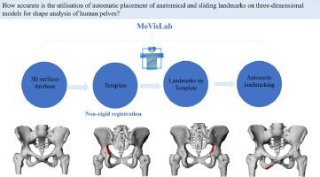
Estimating sex from unknown human skeletal remains is an important component in forensic anthropology. Currently, both morphological and morphometric methods are used for sex estimation. These methods employ landmarks to make morphological comparisons between and within groups. Manual landmarking has been regarded as time-consuming and subjective. To decrease observer subjectivity and reduce measurement errors, an automated three-dimensional (3D) method was developed. This study aimed to validate the utilisation of the automatic placement of anatomical and sliding landmarks on 3D pelvis models for shape analysis using Computed Tomography (CT) scans.
Computed Tomography scans of adult South Africans were obtained from Steve Biko Academic Hospital, Pretoria, South Africa. In this study, automatic landmarking was validated on 130 3D reconstructions of the adult human pelvis. Eighteen anatomical and 260 sliding landmarks were registered on 130 3D models of the same individuals manually and automatically using the MeVisLab © v 2.7.1 software. Landmark datasets were acquired using both landmarking methods and compared using reproducibility testing and geometric morphometric (GMM) analysis. Reproducibility testing of both landmark datasets demonstrated minimal dispersion errors (<2 mm), indicating the reliability and repeatability of both landmarking methods. Variance analysis showed that pelvis shape sex-related variation was statistically significant (p <0.05) using both methods. In addition, cross-validated discriminant function analysis (DFA) yielded accuracies between 82.98 – 97.73% and 65.91 – 93.18% using automatic and manual placement of landmarks, respectively.
In forensics using 3D automatic approaches, and advanced statistical analysis might allow forensic anthropologists to estimate sex in a more accurate and repeatable way.
在人体骨盆形状分析的三维模型上自动放置解剖和滑动标志的验证
从未知的人类骨骼遗骸中估计性别是法医人类学的一个重要组成部分。目前,形态学和形态计量学方法都用于性别估计。这些方法使用界标在组之间和组内进行形态学比较。人工土地标记被认为是耗时和主观的。为了减少观察者的主观性并减少测量误差,开发了一种自动三维(3D)方法。本研究旨在验证使用计算机断层扫描(CT)在三维骨盆模型上自动放置解剖和滑动标志进行形状分析的可行性。南非成年人的计算机断层扫描是从南非比勒陀利亚的Steve Biko学术医院获得的。在这项研究中,自动陆地标记在130个成人骨盆的3D重建上得到了验证。使用MeVisLab©v 2.7.1软件手动和自动在130个相同个体的3D模型上注册了18个解剖标志和260个滑动标志。使用两种陆地标记方法获取陆地标记数据集,并使用再现性测试和几何形态计量学(GMM)分析进行比较。两个标志性数据集的再现性测试都证明了最小的分散误差(<;2 mm),表明了两种标志性方法的可靠性和可重复性。方差分析显示,使用这两种方法,骨盆形状性别相关的变异具有统计学意义(p<0.05)。此外,使用自动和手动放置界标,交叉验证判别函数分析(DFA)的准确率分别在82.98–97.73%和65.91–93.18%之间。在使用3D自动方法的法医学中,先进的统计分析可能使法医人类学家能够以更准确和可重复的方式估计性别。
本文章由计算机程序翻译,如有差异,请以英文原文为准。


