Dean Karaica, Ivan Mihaljević, Lana Vujica, Arvena Bošnjak, Jelena Dragojević, Cecile Otten, Nency Babić, Jovica Lončar, Tvrtko Smital
{"title":"Stage-dependent localization of F-actin and Na+/K+-ATPase in zebrafish embryos detected using optimized cryosectioning immunostaining protocol","authors":"Dean Karaica, Ivan Mihaljević, Lana Vujica, Arvena Bošnjak, Jelena Dragojević, Cecile Otten, Nency Babić, Jovica Lončar, Tvrtko Smital","doi":"10.1002/jemt.24270","DOIUrl":null,"url":null,"abstract":"<div>\n \n \n <section>\n \n <p>The increasing use of the zebrafish model in biomedical and (eco)toxicological studies aimed at understanding the function of various proteins highlight the importance of optimizing existing methods to study gene and protein expression and localization in this model. In this context, zebrafish cryosections are still underutilized compared with whole-mount preparations. In this study, we used zebrafish embryos (24–120 hpf) to determine key factors for the preparation of high-quality zebrafish cryosections and to determine the optimal protocol for (immuno)fluorescence analyses of Na<sup>+</sup>/K<sup>+</sup>-ATPase and F-actin, across developmental stages from 1 to 5 dpf. The results showed that the highest quality zebrafish cryosections were obtained after the samples were fixed in 4% paraformaldehyde (PFA) for 1 h, incubated in 2.5% bovine gelatin/25% sucrose mixture, embedded in OCT, and then sectioned to 8 μm thickness at −20°C. Fluorescence microscopy analysis of phalloidin-labeled zebrafish skeletal muscle revealed that 1-h-4% PFA-fixed samples allowed optimal binding of phalloidin to F-actin. Further immunofluorescence analyses revealed detailed localization of F-actin and Na<sup>+</sup>/K<sup>+</sup>-ATPase in various tissues of the zebrafish and a stage-dependent increase in their respective expression in the somitic muscles and pronephros. Finally, staining of zebrafish cryosections and whole-mount samples revealed organ-specific and zone-dependent localizations of the Na<sup>+</sup>/K<sup>+</sup>-ATPase α1-subunit.</p>\n </section>\n \n <section>\n \n <h3> Research Highlights</h3>\n \n <p>This study brings optimization of existing protocols for preparation and use of zebrafish embryos cryosections in (immuno)histological analyses. It reveals stage-dependent localization/expression of F-actin and Na<sup>+</sup>/K<sup>+</sup>-ATPase in zebrafish embryos.</p>\n </section>\n </div>","PeriodicalId":18684,"journal":{"name":"Microscopy Research and Technique","volume":"86 3","pages":"294-310"},"PeriodicalIF":2.1000,"publicationDate":"2022-12-01","publicationTypes":"Journal Article","fieldsOfStudy":null,"isOpenAccess":false,"openAccessPdf":"","citationCount":"0","resultStr":null,"platform":"Semanticscholar","paperid":null,"PeriodicalName":"Microscopy Research and Technique","FirstCategoryId":"5","ListUrlMain":"https://onlinelibrary.wiley.com/doi/10.1002/jemt.24270","RegionNum":3,"RegionCategory":"工程技术","ArticlePicture":[],"TitleCN":null,"AbstractTextCN":null,"PMCID":null,"EPubDate":"","PubModel":"","JCR":"Q2","JCRName":"ANATOMY & MORPHOLOGY","Score":null,"Total":0}
引用次数: 0
Abstract
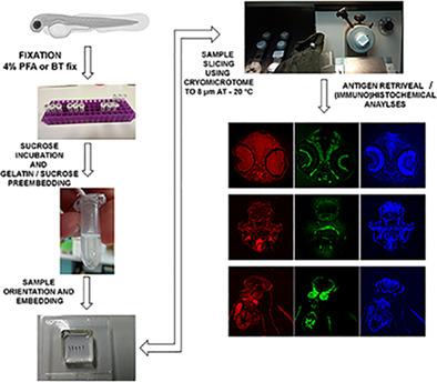
The increasing use of the zebrafish model in biomedical and (eco)toxicological studies aimed at understanding the function of various proteins highlight the importance of optimizing existing methods to study gene and protein expression and localization in this model. In this context, zebrafish cryosections are still underutilized compared with whole-mount preparations. In this study, we used zebrafish embryos (24–120 hpf) to determine key factors for the preparation of high-quality zebrafish cryosections and to determine the optimal protocol for (immuno)fluorescence analyses of Na+/K+-ATPase and F-actin, across developmental stages from 1 to 5 dpf. The results showed that the highest quality zebrafish cryosections were obtained after the samples were fixed in 4% paraformaldehyde (PFA) for 1 h, incubated in 2.5% bovine gelatin/25% sucrose mixture, embedded in OCT, and then sectioned to 8 μm thickness at −20°C. Fluorescence microscopy analysis of phalloidin-labeled zebrafish skeletal muscle revealed that 1-h-4% PFA-fixed samples allowed optimal binding of phalloidin to F-actin. Further immunofluorescence analyses revealed detailed localization of F-actin and Na+/K+-ATPase in various tissues of the zebrafish and a stage-dependent increase in their respective expression in the somitic muscles and pronephros. Finally, staining of zebrafish cryosections and whole-mount samples revealed organ-specific and zone-dependent localizations of the Na+/K+-ATPase α1-subunit.
Research Highlights
This study brings optimization of existing protocols for preparation and use of zebrafish embryos cryosections in (immuno)histological analyses. It reveals stage-dependent localization/expression of F-actin and Na+/K+-ATPase in zebrafish embryos.
期刊介绍:
Microscopy Research and Technique (MRT) publishes articles on all aspects of advanced microscopy original architecture and methodologies with applications in the biological, clinical, chemical, and materials sciences. Original basic and applied research as well as technical papers dealing with the various subsets of microscopy are encouraged. MRT is the right form for those developing new microscopy methods or using the microscope to answer key questions in basic and applied research.


 分享
分享
 求助内容:
求助内容: 应助结果提醒方式:
 扫码关注我们
