{"title":"Microstructural evolution of tantalum nitride thin films synthesized by inductively coupled plasma sputtering","authors":"Sung-Il Baik, Young-Woon Kim","doi":"10.1186/s42649-020-00026-7","DOIUrl":null,"url":null,"abstract":"<p>Tantalum nitride (TaN<sub>x</sub>) thin films were grown utilizing an inductively coupled plasma (ICP) assisted direct current (DC) sputtering, and 20–100% improved microhardness values were obtained. The detailed microstructural changes of the TaN<sub>x</sub> films were characterized utilizing transmission electron microscopy (TEM), as a function of nitrogen gas fraction and ICP power. As nitrogen gas fraction increases?from 0.05 to 0.15, the TaN<sub>x</sub> phase evolves from body-centered-cubic (b.c.c.) TaN<sub>0.1</sub>, to face-centered-cubic (f.c.c.) δ-TaN, to hexagonal-close-packing (h.c.p.) ε-TaN phase. By increasing ICP power from 100?W to 400?W, the f.c.c. δ- TaN phase becomes the main phase in all nitrogen fractions investigated. The higher ICP power enhances the mobility of Ta and N ions, which stabilizes the δ-TaN phase like a high-temperature regime and removes the micro-voids between the columnar grains in the TaN<sub>x</sub> film. The dense δ-TaN structure with reduced columnar grains and micro-voids increases the strength of the TaN<sub>x</sub> film.</p>","PeriodicalId":470,"journal":{"name":"Applied Microscopy","volume":"50 1","pages":""},"PeriodicalIF":0.0000,"publicationDate":"2020-02-27","publicationTypes":"Journal Article","fieldsOfStudy":null,"isOpenAccess":false,"openAccessPdf":"https://sci-hub-pdf.com/10.1186/s42649-020-00026-7","citationCount":"4","resultStr":null,"platform":"Semanticscholar","paperid":null,"PeriodicalName":"Applied Microscopy","FirstCategoryId":"1085","ListUrlMain":"https://link.springer.com/article/10.1186/s42649-020-00026-7","RegionNum":0,"RegionCategory":null,"ArticlePicture":[],"TitleCN":null,"AbstractTextCN":null,"PMCID":null,"EPubDate":"","PubModel":"","JCR":"Q3","JCRName":"Immunology and Microbiology","Score":null,"Total":0}
引用次数: 4
Abstract
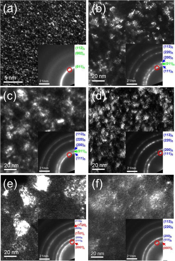
Tantalum nitride (TaNx) thin films were grown utilizing an inductively coupled plasma (ICP) assisted direct current (DC) sputtering, and 20–100% improved microhardness values were obtained. The detailed microstructural changes of the TaNx films were characterized utilizing transmission electron microscopy (TEM), as a function of nitrogen gas fraction and ICP power. As nitrogen gas fraction increases?from 0.05 to 0.15, the TaNx phase evolves from body-centered-cubic (b.c.c.) TaN0.1, to face-centered-cubic (f.c.c.) δ-TaN, to hexagonal-close-packing (h.c.p.) ε-TaN phase. By increasing ICP power from 100?W to 400?W, the f.c.c. δ- TaN phase becomes the main phase in all nitrogen fractions investigated. The higher ICP power enhances the mobility of Ta and N ions, which stabilizes the δ-TaN phase like a high-temperature regime and removes the micro-voids between the columnar grains in the TaNx film. The dense δ-TaN structure with reduced columnar grains and micro-voids increases the strength of the TaNx film.
Applied MicroscopyImmunology and Microbiology-Applied Microbiology and Biotechnology
CiteScore
3.40
自引率
0.00%
发文量
10
审稿时长
10 weeks
期刊介绍:
Applied Microscopy is a peer-reviewed journal sponsored by the Korean Society of Microscopy. The journal covers all the interdisciplinary fields of technological developments in new microscopy methods and instrumentation and their applications to biological or materials science for determining structure and chemistry. ISSN: 22875123, 22874445.


 分享
分享
 求助内容:
求助内容: 应助结果提醒方式:
 扫码关注我们
