Yuki Toyama, Atul Kaushik Rangadurai, Lewis E. Kay
{"title":"Measurement of 1Hα transverse relaxation rates in proteins: application to solvent PREs","authors":"Yuki Toyama, Atul Kaushik Rangadurai, Lewis E. Kay","doi":"10.1007/s10858-022-00401-4","DOIUrl":null,"url":null,"abstract":"<div><p>It has recently been demonstrated that accurate near surface electrostatic potentials can be calculated for proteins from solvent paramagnetic relaxation enhancements (PREs) of amide protons measured using spin labels of similar structures but different charges (Yu <i>et al</i>. in Proc Natl Acad Sci 118(25):e2104020118, 2021). Here we develop methodology for extending such measurements to intrinsically disordered proteins at neutral pH where amide spectra are of very poor quality. Under these conditions it is shown that accurate PRE values can be measured using the haCONHA experiment that has been modified for recording <sup>1</sup>H<sup>α</sup> transverse relaxation rates. The optimal pulse scheme includes a spin-lock relaxation element for suppression of homonuclear scalar coupled evolution for all <sup>1</sup>H<sup>α</sup> protons, except those derived from Ser and Thr residues, and minimizes the radiation damping field from water magnetization that would otherwise increase measured relaxation rates. The robustness of the experiment is verified by developing a second approach using a band selective adiabatic decoupling scheme for suppression of scalar coupling modulations during <sup>1</sup>H<sup>α</sup> relaxation and showing that the measured PRE values from the two methods are in excellent agreement. The near surface electrostatic potential of a 103-residue construct comprising the C-terminal intrinsically disordered region of the RNA-binding protein CAPRIN1 is obtained at pH 5.5 using both <sup>1</sup>H<sup>N</sup> and <sup>1</sup>H<sup>α</sup>-based relaxation rates, and at pH 7.4 where only <sup>1</sup>H<sup>α</sup> rates can be quantified, with very good agreement between potentials obtained under all experimental conditions.\n</p></div>","PeriodicalId":613,"journal":{"name":"Journal of Biomolecular NMR","volume":"76 4","pages":"137 - 152"},"PeriodicalIF":1.9000,"publicationDate":"2022-08-26","publicationTypes":"Journal Article","fieldsOfStudy":null,"isOpenAccess":false,"openAccessPdf":"","citationCount":"7","resultStr":null,"platform":"Semanticscholar","paperid":null,"PeriodicalName":"Journal of Biomolecular NMR","FirstCategoryId":"99","ListUrlMain":"https://link.springer.com/article/10.1007/s10858-022-00401-4","RegionNum":3,"RegionCategory":"生物学","ArticlePicture":[],"TitleCN":null,"AbstractTextCN":null,"PMCID":null,"EPubDate":"","PubModel":"","JCR":"Q3","JCRName":"BIOCHEMISTRY & MOLECULAR BIOLOGY","Score":null,"Total":0}
引用次数: 7
Abstract
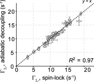
It has recently been demonstrated that accurate near surface electrostatic potentials can be calculated for proteins from solvent paramagnetic relaxation enhancements (PREs) of amide protons measured using spin labels of similar structures but different charges (Yu et al. in Proc Natl Acad Sci 118(25):e2104020118, 2021). Here we develop methodology for extending such measurements to intrinsically disordered proteins at neutral pH where amide spectra are of very poor quality. Under these conditions it is shown that accurate PRE values can be measured using the haCONHA experiment that has been modified for recording 1Hα transverse relaxation rates. The optimal pulse scheme includes a spin-lock relaxation element for suppression of homonuclear scalar coupled evolution for all 1Hα protons, except those derived from Ser and Thr residues, and minimizes the radiation damping field from water magnetization that would otherwise increase measured relaxation rates. The robustness of the experiment is verified by developing a second approach using a band selective adiabatic decoupling scheme for suppression of scalar coupling modulations during 1Hα relaxation and showing that the measured PRE values from the two methods are in excellent agreement. The near surface electrostatic potential of a 103-residue construct comprising the C-terminal intrinsically disordered region of the RNA-binding protein CAPRIN1 is obtained at pH 5.5 using both 1HN and 1Hα-based relaxation rates, and at pH 7.4 where only 1Hα rates can be quantified, with very good agreement between potentials obtained under all experimental conditions.
最近有研究表明,通过使用结构相似但电荷不同的自旋标签测量酰胺质子的溶剂顺磁弛豫增强(PREs),可以计算出蛋白质的精确近表面静电电位(Yu et al. in Proc Natl Acad Sci 118(25):e2104020118, 2021)。在这里,我们开发了一种方法,可以将这种测量扩展到中性pH下的内在无序蛋白质,其中酰胺光谱的质量非常差。在这些条件下,使用经过修改的haCONHA实验可以测量精确的PRE值,以记录1Hα横向弛豫速率。最佳脉冲方案包括一个自旋锁弛豫元件,用于抑制所有1Hα质子的同核标量耦合演化,除了来自Ser和Thr残基的质子,并且最小化水磁化的辐射阻尼场,否则会增加测量的弛豫率。通过采用带选择性绝热解耦方案来抑制1Hα弛豫期间的标量耦合调制,验证了实验的鲁棒性,并表明两种方法测量的PRE值非常一致。包含rna结合蛋白CAPRIN1的c端固有无序区的103个残基结构体的近表面静电电位在pH为5.5时使用1HN和1Hα为基础的弛豫速率,在pH为7.4时仅可以量化1Hα速率,在所有实验条件下获得的电位之间具有非常好的一致性。
期刊介绍:
The Journal of Biomolecular NMR provides a forum for publishing research on technical developments and innovative applications of nuclear magnetic resonance spectroscopy for the study of structure and dynamic properties of biopolymers in solution, liquid crystals, solids and mixed environments, e.g., attached to membranes. This may include:
Three-dimensional structure determination of biological macromolecules (polypeptides/proteins, DNA, RNA, oligosaccharides) by NMR.
New NMR techniques for studies of biological macromolecules.
Novel approaches to computer-aided automated analysis of multidimensional NMR spectra.
Computational methods for the structural interpretation of NMR data, including structure refinement.
Comparisons of structures determined by NMR with those obtained by other methods, e.g. by diffraction techniques with protein single crystals.
New techniques of sample preparation for NMR experiments (biosynthetic and chemical methods for isotope labeling, preparation of nutrients for biosynthetic isotope labeling, etc.). An NMR characterization of the products must be included.


 分享
分享
 求助内容:
求助内容: 应助结果提醒方式:
 扫码关注我们
