Zahra Vasegh, Yaser Safi, Maryam Sanaei Azar, Mitra Ghazizadeh Ahsaie, S Marjan Arianezhad
{"title":"Assessment of bony changes in temporomandibular joint in patients using cone beam computed tomography - a cross sectional study.","authors":"Zahra Vasegh, Yaser Safi, Maryam Sanaei Azar, Mitra Ghazizadeh Ahsaie, S Marjan Arianezhad","doi":"10.1186/s13005-023-00392-z","DOIUrl":null,"url":null,"abstract":"<p><strong>Background and aim: </strong>The aim of this study is to evaluate the changes in the temporomandibular joint (TMJ) in patients with temporomandibular disorder (TMD) and the relationship between age, sex, and types of TMJ change using Cone Beam Computed Tomography (CBCT).</p><p><strong>Methods and material: </strong>CBCT records of 200 patients (123 women and 67 men) were retrieved and assessed. Right and left TMJs were evaluated separately, resulting in a total of 400 TMJs. The images were analyzed using On demand 3D Application The radiographic findings were classified as erosion, proliferative changes mainly, including flattening and osteophytes of the condyle, sclerosis, Ely cyst, hypoplasia and hyperplasia of the condyles, ankylosis, and joint cavity. Data analysis was performed using descriptive statistics, paired T-tests, and repeated measure ANOVA (Analysis of Variance) in SPSS Software.</p><p><strong>Results: </strong>The most prevalent types of condylar bony changes observed was osteophyte (63.5%) followed by flattening of the articular surface (42%), erosion (40%), ankylosis (10%) and sclerosis (10%). 7.5% of joints showed hyperplastic condyles but only 2% showed hypoplasia. The least prevalent change observed was Ely Cyst (1%). Osteophyte was the most prevalent change observed in all age groups and both sexes except for men aged 31 ~ 50, where flattening was more frequent. A statistically significant difference was found between sex and prevalence of erosion in the age group of 10 ~ 30 (P = 0.001); as well as between sex and condylar hyperplasia in the same age group.</p><p><strong>Conclusion: </strong>Based on the findings of this research, the prevalence of bony changes of TMJ from highest to lowest is as follows: osteophyte, flattening of the articular surface, erosion, ankylosis, sclerosis, hyperplastic condyles, hypoplastic condyles and Ely Cyst. CBCT is an accurate 3 dimensional imaging modality for assessment of TMJ bony structures.</p>","PeriodicalId":12994,"journal":{"name":"Head & Face Medicine","volume":"19 1","pages":"47"},"PeriodicalIF":2.4000,"publicationDate":"2023-10-28","publicationTypes":"Journal Article","fieldsOfStudy":null,"isOpenAccess":false,"openAccessPdf":"https://www.ncbi.nlm.nih.gov/pmc/articles/PMC10612346/pdf/","citationCount":"0","resultStr":null,"platform":"Semanticscholar","paperid":null,"PeriodicalName":"Head & Face Medicine","FirstCategoryId":"3","ListUrlMain":"https://doi.org/10.1186/s13005-023-00392-z","RegionNum":2,"RegionCategory":"医学","ArticlePicture":[],"TitleCN":null,"AbstractTextCN":null,"PMCID":null,"EPubDate":"","PubModel":"","JCR":"Q2","JCRName":"DENTISTRY, ORAL SURGERY & MEDICINE","Score":null,"Total":0}
引用次数: 0
Abstract
Background and aim: The aim of this study is to evaluate the changes in the temporomandibular joint (TMJ) in patients with temporomandibular disorder (TMD) and the relationship between age, sex, and types of TMJ change using Cone Beam Computed Tomography (CBCT).
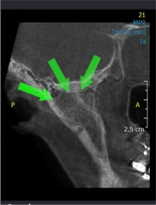
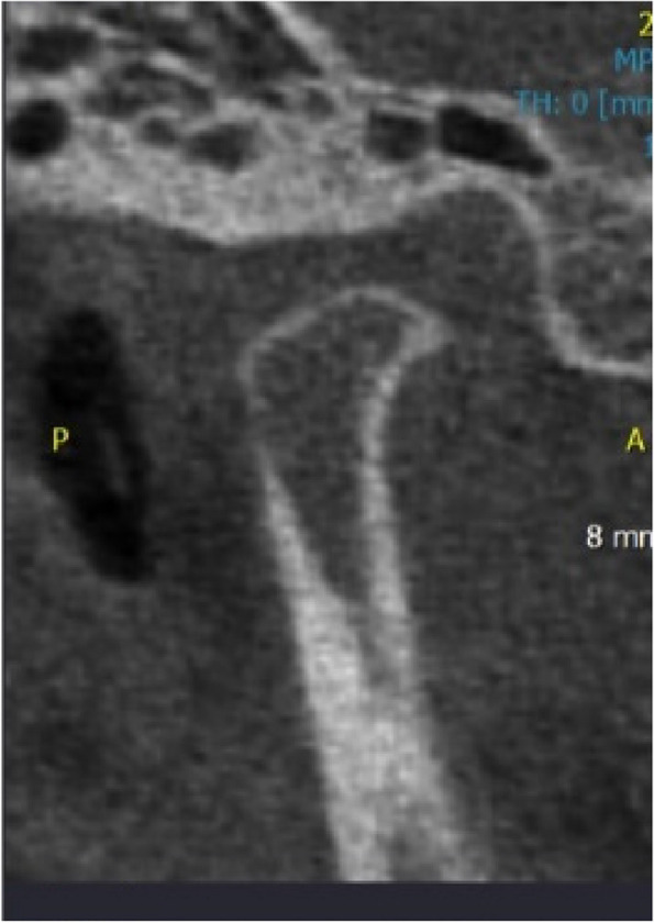
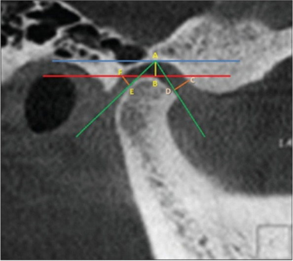
Methods and material: CBCT records of 200 patients (123 women and 67 men) were retrieved and assessed. Right and left TMJs were evaluated separately, resulting in a total of 400 TMJs. The images were analyzed using On demand 3D Application The radiographic findings were classified as erosion, proliferative changes mainly, including flattening and osteophytes of the condyle, sclerosis, Ely cyst, hypoplasia and hyperplasia of the condyles, ankylosis, and joint cavity. Data analysis was performed using descriptive statistics, paired T-tests, and repeated measure ANOVA (Analysis of Variance) in SPSS Software.
Results: The most prevalent types of condylar bony changes observed was osteophyte (63.5%) followed by flattening of the articular surface (42%), erosion (40%), ankylosis (10%) and sclerosis (10%). 7.5% of joints showed hyperplastic condyles but only 2% showed hypoplasia. The least prevalent change observed was Ely Cyst (1%). Osteophyte was the most prevalent change observed in all age groups and both sexes except for men aged 31 ~ 50, where flattening was more frequent. A statistically significant difference was found between sex and prevalence of erosion in the age group of 10 ~ 30 (P = 0.001); as well as between sex and condylar hyperplasia in the same age group.
Conclusion: Based on the findings of this research, the prevalence of bony changes of TMJ from highest to lowest is as follows: osteophyte, flattening of the articular surface, erosion, ankylosis, sclerosis, hyperplastic condyles, hypoplastic condyles and Ely Cyst. CBCT is an accurate 3 dimensional imaging modality for assessment of TMJ bony structures.
期刊介绍:
Head & Face Medicine is a multidisciplinary open access journal that publishes basic and clinical research concerning all aspects of cranial, facial and oral conditions.
The journal covers all aspects of cranial, facial and oral diseases and their management. It has been designed as a multidisciplinary journal for clinicians and researchers involved in the diagnostic and therapeutic aspects of diseases which affect the human head and face. The journal is wide-ranging, covering the development, aetiology, epidemiology and therapy of head and face diseases to the basic science that underlies these diseases. Management of head and face diseases includes all aspects of surgical and non-surgical treatments including psychopharmacological therapies.


 分享
分享
 求助内容:
求助内容: 应助结果提醒方式:
 扫码关注我们
