Anthony Yan-Tang Wu, Ponarulselvam Sekar, Duen-Yi Huang, Shu-Hao Hsu, Chi-Ming Chan, Wan-Wan Lin
{"title":"Spatiotemporal roles of AMPK in PARP-1- and autophagy-dependent retinal pigment epithelial cell death caused by UVA.","authors":"Anthony Yan-Tang Wu, Ponarulselvam Sekar, Duen-Yi Huang, Shu-Hao Hsu, Chi-Ming Chan, Wan-Wan Lin","doi":"10.1186/s12929-023-00978-4","DOIUrl":null,"url":null,"abstract":"<p><strong>Background: </strong>Although stimulating autophagy caused by UV has been widely demonstrated in skin cells to exert cell protection, it remains unknown the cellular events in UVA-treated retinal pigment epithelial (RPE) cells.</p><p><strong>Methods: </strong>Human ARPE-19 cells were used to measure cell viability, mitochondrial reactive oxygen species (ROS), mitochondrial membrane potential (MMP), mitochondrial mass and lysosomal mass by flow cytometry. Mitochondrial oxygen consumption rate (OCR) was recorded using Seahorse XF flux analyzer. Confocal microscopic images were performed to indicate the mitochondrial dynamics, LC3 level, and AMPK translocation after UVA irradiation.</p><p><strong>Results: </strong>We confirmed mitochondrial ROS production and DNA damage are two major features caused by UVA. We found the cell death is prevented by autophagy inhibitor 3-methyladenine and gene silencing of ATG5, and UVA induces ROS-dependent LC3II expression, LC3 punctate and TFEB expression, suggesting the autophagic death in the UVA-stressed RPE cells. Although PARP-1 inhibitor olaparib increases DNA damage, ROS production, and cell death, it also blocks AMPK activation caused by UVA. Interestingly we found a dramatic nuclear export of AMPK upon UVA irradiation which is blocked by N-acetylcysteine and olaparib. In addition, UVA exposure gradually decreases lysosomal mass and inhibits cathepsin B activity at late phase due to lysosomal dysfunction. Nevertheless, cathepsin B inhibitor, CA-074Me, reverses the death extent, suggesting the contribution of cathepsin B in the death pathway. When examining the role of EGFR in cellular events caused by UVA, we found that UVA can rapidly transactivate EGFR, and treatment with EGFR TKIs (gefitinib and afatinib) enhances the cell death accompanied by the increased LC3II formation, ROS production, loss of MMP and mass of mitochondria and lysosomes. Although AMPK activation by ROS-PARP-1 mediates autophagic cell death, we surprisingly found that pretreatment of cells with AMPK activators (A769662 and metformin) reverses cell death. Concomitantly, both agents block UVA-induced mitochondrial ROS production, autophagic flux, and mitochondrial fission without changing the inhibition of cathepsin B.</p><p><strong>Conclusion: </strong>UVA exposure rapidly induces ROS-PARP-1-AMPK-autophagic flux and late lysosomal dysfunction. Pre-inducing AMPK activation can prevent cellular events caused by UVA and provide a new protective strategy in photo-oxidative stress and photo-retinopathy.</p>","PeriodicalId":15365,"journal":{"name":"Journal of Biomedical Science","volume":"30 1","pages":"91"},"PeriodicalIF":12.1000,"publicationDate":"2023-11-07","publicationTypes":"Journal Article","fieldsOfStudy":null,"isOpenAccess":false,"openAccessPdf":"https://www.ncbi.nlm.nih.gov/pmc/articles/PMC10629085/pdf/","citationCount":"0","resultStr":null,"platform":"Semanticscholar","paperid":null,"PeriodicalName":"Journal of Biomedical Science","FirstCategoryId":"3","ListUrlMain":"https://doi.org/10.1186/s12929-023-00978-4","RegionNum":2,"RegionCategory":"医学","ArticlePicture":[],"TitleCN":null,"AbstractTextCN":null,"PMCID":null,"EPubDate":"","PubModel":"","JCR":"Q1","JCRName":"CELL BIOLOGY","Score":null,"Total":0}
引用次数: 0
Abstract
Background: Although stimulating autophagy caused by UV has been widely demonstrated in skin cells to exert cell protection, it remains unknown the cellular events in UVA-treated retinal pigment epithelial (RPE) cells.
Methods: Human ARPE-19 cells were used to measure cell viability, mitochondrial reactive oxygen species (ROS), mitochondrial membrane potential (MMP), mitochondrial mass and lysosomal mass by flow cytometry. Mitochondrial oxygen consumption rate (OCR) was recorded using Seahorse XF flux analyzer. Confocal microscopic images were performed to indicate the mitochondrial dynamics, LC3 level, and AMPK translocation after UVA irradiation.
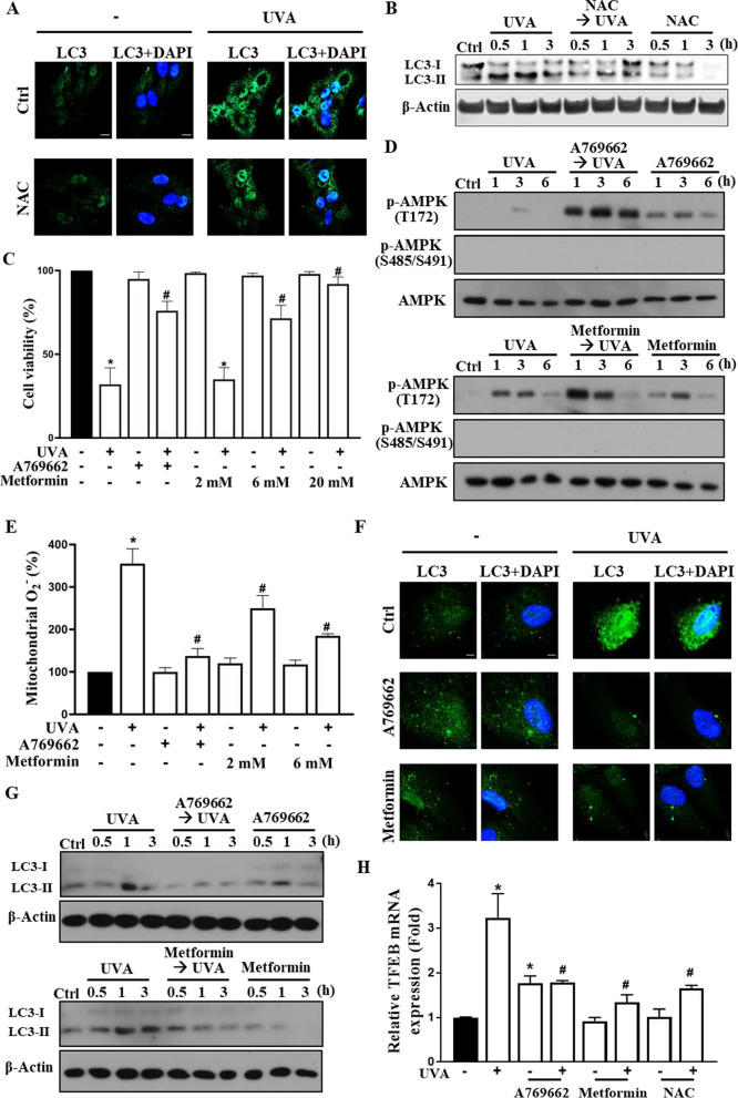
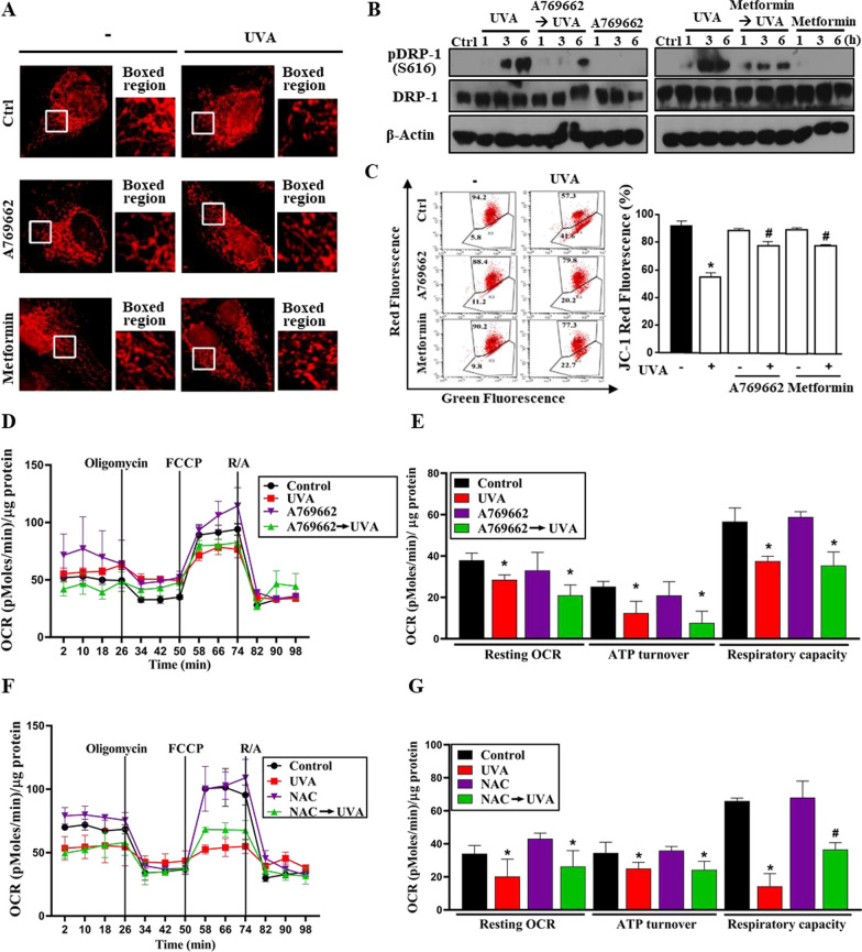
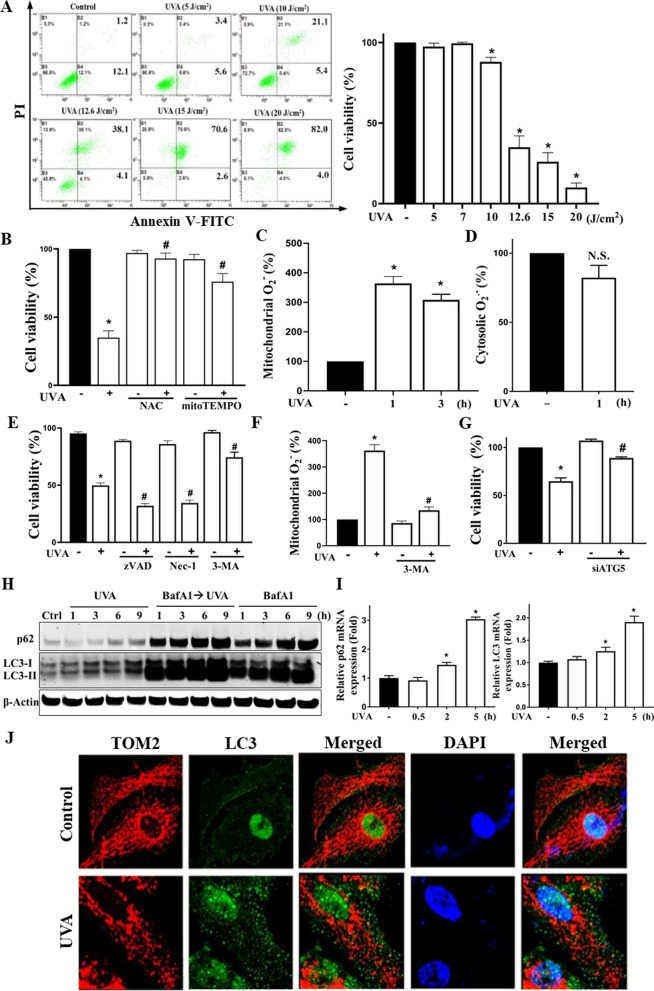
Results: We confirmed mitochondrial ROS production and DNA damage are two major features caused by UVA. We found the cell death is prevented by autophagy inhibitor 3-methyladenine and gene silencing of ATG5, and UVA induces ROS-dependent LC3II expression, LC3 punctate and TFEB expression, suggesting the autophagic death in the UVA-stressed RPE cells. Although PARP-1 inhibitor olaparib increases DNA damage, ROS production, and cell death, it also blocks AMPK activation caused by UVA. Interestingly we found a dramatic nuclear export of AMPK upon UVA irradiation which is blocked by N-acetylcysteine and olaparib. In addition, UVA exposure gradually decreases lysosomal mass and inhibits cathepsin B activity at late phase due to lysosomal dysfunction. Nevertheless, cathepsin B inhibitor, CA-074Me, reverses the death extent, suggesting the contribution of cathepsin B in the death pathway. When examining the role of EGFR in cellular events caused by UVA, we found that UVA can rapidly transactivate EGFR, and treatment with EGFR TKIs (gefitinib and afatinib) enhances the cell death accompanied by the increased LC3II formation, ROS production, loss of MMP and mass of mitochondria and lysosomes. Although AMPK activation by ROS-PARP-1 mediates autophagic cell death, we surprisingly found that pretreatment of cells with AMPK activators (A769662 and metformin) reverses cell death. Concomitantly, both agents block UVA-induced mitochondrial ROS production, autophagic flux, and mitochondrial fission without changing the inhibition of cathepsin B.
Conclusion: UVA exposure rapidly induces ROS-PARP-1-AMPK-autophagic flux and late lysosomal dysfunction. Pre-inducing AMPK activation can prevent cellular events caused by UVA and provide a new protective strategy in photo-oxidative stress and photo-retinopathy.
期刊介绍:
The Journal of Biomedical Science is an open access, peer-reviewed journal that focuses on fundamental and molecular aspects of basic medical sciences. It emphasizes molecular studies of biomedical problems and mechanisms. The National Science and Technology Council (NSTC), Taiwan supports the journal and covers the publication costs for accepted articles. The journal aims to provide an international platform for interdisciplinary discussions and contribute to the advancement of medicine. It benefits both readers and authors by accelerating the dissemination of research information and providing maximum access to scholarly communication. All articles published in the Journal of Biomedical Science are included in various databases such as Biological Abstracts, BIOSIS, CABI, CAS, Citebase, Current contents, DOAJ, Embase, EmBiology, and Global Health, among others.


 分享
分享
 求助内容:
求助内容: 应助结果提醒方式:
 扫码关注我们
