Mario Fernando Ortega-Mafla, Valerye Viveros-Gonzalez, Wilmar Saldarriaga-Gil
{"title":"Thoracic kidney and diaphragmatic hernia: prenatal diagnosis and successful management. Case report and review of the literature","authors":"Mario Fernando Ortega-Mafla, Valerye Viveros-Gonzalez, Wilmar Saldarriaga-Gil","doi":"10.18597/rcog.4020","DOIUrl":null,"url":null,"abstract":"<p><strong>Objectives: </strong>To report a case of prenatal diagnosis of ectopic intrathoracic kidney with diaphragmatic hernia managed surgically after birth, and to conduct a review of the literature on prenatal diagnosis of ectopic intrathoracic kidney and perinatal prognosis.</p><p><strong>Material and methods: </strong>We report the case of a 28-week fetus in which, on ultrasound imaging, a mass was observed displacing the heart and lung in the right hemithorax, which was was confirmed by magnetic resonance (MR) to be an ectopic intrathoracic kidney (ITEK). After birth, the neonate was approached by laparoscopy to place a mesh in continuity with the diaphragm, leaving the kidney in the abdomen, with good evolution. A search was conducted in the PubMed, Embase and Cochrane databases for cohorts, case reports and case series of prenatal diagnosis of intrathoracic kidney in the fetus. Information was retrieved regarding design, population, imaging diagnosis, treatment and prognosis.</p><p><strong>Results: </strong>The search identified 8 studies that met the inclusion criteria, reporting a total of 8 cases. Ultrasound diagnosis showed ectopic intrathoracic kidney associated with diaphragmatic hernia in all the subjects. Fetal magnetic resonance imaging (MRI) was also used in 5 cases.</p><p><strong>Conclusions: </strong>Ectopic intrathoracic kidney is a congenital abnormality amenable to prenatal diagnosis. Survival after corrective surgery performed in the neonatal period is common. There is a paucity of publications, limited to case reports, regarding the prenatal diagnosis of this condition.</p>","PeriodicalId":101422,"journal":{"name":"Revista colombiana de obstetricia y ginecologia","volume":"74 3","pages":"237-243"},"PeriodicalIF":0.0000,"publicationDate":"2023-09-30","publicationTypes":"Journal Article","fieldsOfStudy":null,"isOpenAccess":false,"openAccessPdf":"https://www.ncbi.nlm.nih.gov/pmc/articles/PMC10652776/pdf/","citationCount":"0","resultStr":null,"platform":"Semanticscholar","paperid":null,"PeriodicalName":"Revista colombiana de obstetricia y ginecologia","FirstCategoryId":"1085","ListUrlMain":"https://doi.org/10.18597/rcog.4020","RegionNum":0,"RegionCategory":null,"ArticlePicture":[],"TitleCN":null,"AbstractTextCN":null,"PMCID":null,"EPubDate":"","PubModel":"","JCR":"","JCRName":"","Score":null,"Total":0}
引用次数: 0
Abstract
Objectives: To report a case of prenatal diagnosis of ectopic intrathoracic kidney with diaphragmatic hernia managed surgically after birth, and to conduct a review of the literature on prenatal diagnosis of ectopic intrathoracic kidney and perinatal prognosis.
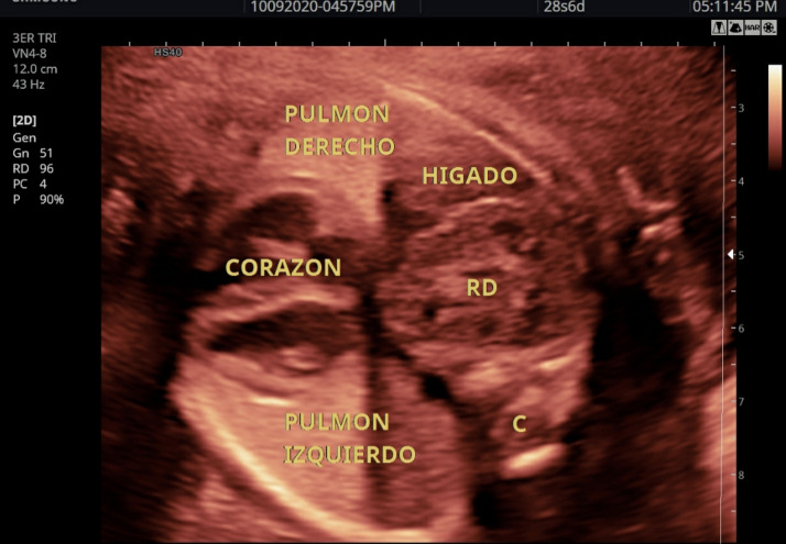
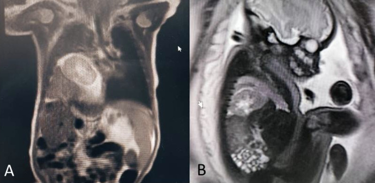
Material and methods: We report the case of a 28-week fetus in which, on ultrasound imaging, a mass was observed displacing the heart and lung in the right hemithorax, which was was confirmed by magnetic resonance (MR) to be an ectopic intrathoracic kidney (ITEK). After birth, the neonate was approached by laparoscopy to place a mesh in continuity with the diaphragm, leaving the kidney in the abdomen, with good evolution. A search was conducted in the PubMed, Embase and Cochrane databases for cohorts, case reports and case series of prenatal diagnosis of intrathoracic kidney in the fetus. Information was retrieved regarding design, population, imaging diagnosis, treatment and prognosis.
Results: The search identified 8 studies that met the inclusion criteria, reporting a total of 8 cases. Ultrasound diagnosis showed ectopic intrathoracic kidney associated with diaphragmatic hernia in all the subjects. Fetal magnetic resonance imaging (MRI) was also used in 5 cases.
Conclusions: Ectopic intrathoracic kidney is a congenital abnormality amenable to prenatal diagnosis. Survival after corrective surgery performed in the neonatal period is common. There is a paucity of publications, limited to case reports, regarding the prenatal diagnosis of this condition.


 分享
分享

 求助内容:
求助内容: 应助结果提醒方式:
 扫码关注我们
