{"title":"M-CSFR expression in the embryonal component of hepatoblastoma and cell-to-cell interaction between macrophages and hepatoblastoma.","authors":"Lianbo Li, Tomoaki Irie, Daiki Yoshii, Yoshihiro Komohara, Yukio Fujiwara, Shigeyuki Esumi, Masashi Kadohisa, Masaki Honda, Shinya Suzu, Toshiharu Matsuura, Kenichi Kohashi, Yoshinao Oda, Taizo Hibi","doi":"10.1007/s00795-022-00323-y","DOIUrl":null,"url":null,"abstract":"<p><p>Tumor-associated macrophages (TAMs) have protumor functions in various cancers. However, their significance in hepatoblastoma, the most common liver tumor in children, remains unclear. The aim of this study was to explore the potential roles of TAMs in hepatoblastoma. Immunohistochemical analysis revealed that the density of CD204-positive TAMs was significantly higher in the embryonal component than in other histological subtypes of hepatoblastoma. An in vitro co-culture study with Huh6 cells and human monocyte-derived macrophages (HMDMs) showed that macrophage-colony-stimulating factor receptor (M-CSFR) was strongly up-regulated in the Huh6 cells that were directly co-cultured with HMDMs. The expressions of M-CSFR ligands (interleukin-34 and M-CSF) were also increased by co-culture with HMDMs. The proliferation of HepG2 cells (another hepatoblastoma cell line expressing M-CSFR) was inhibited by an M-CSFR inhibitor. M-CSFR was found to be highly expressed in the embryonal component and in recurrent lesions. The number of CD204-positive macrophages was also higher in the M-CSFR-positive areas than in the M-CSFR-negative areas. Thus, M-CSFR expression appeared to be induced by cell-cell contact with macrophages in hepatoblastoma cells, and M-CSFR inhibitor is potentially effective against M-CSFR-positive hepatoblastoma, especially recurrent cases.</p>","PeriodicalId":18338,"journal":{"name":"Medical Molecular Morphology","volume":"16 1","pages":"236-247"},"PeriodicalIF":1.1000,"publicationDate":"2022-09-01","publicationTypes":"Journal Article","fieldsOfStudy":null,"isOpenAccess":false,"openAccessPdf":"","citationCount":"0","resultStr":null,"platform":"Semanticscholar","paperid":null,"PeriodicalName":"Medical Molecular Morphology","FirstCategoryId":"3","ListUrlMain":"https://doi.org/10.1007/s00795-022-00323-y","RegionNum":4,"RegionCategory":"医学","ArticlePicture":[],"TitleCN":null,"AbstractTextCN":null,"PMCID":null,"EPubDate":"2022/5/21 0:00:00","PubModel":"Epub","JCR":"Q3","JCRName":"PATHOLOGY","Score":null,"Total":0}
引用次数: 0
Abstract
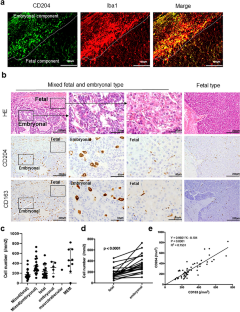
Tumor-associated macrophages (TAMs) have protumor functions in various cancers. However, their significance in hepatoblastoma, the most common liver tumor in children, remains unclear. The aim of this study was to explore the potential roles of TAMs in hepatoblastoma. Immunohistochemical analysis revealed that the density of CD204-positive TAMs was significantly higher in the embryonal component than in other histological subtypes of hepatoblastoma. An in vitro co-culture study with Huh6 cells and human monocyte-derived macrophages (HMDMs) showed that macrophage-colony-stimulating factor receptor (M-CSFR) was strongly up-regulated in the Huh6 cells that were directly co-cultured with HMDMs. The expressions of M-CSFR ligands (interleukin-34 and M-CSF) were also increased by co-culture with HMDMs. The proliferation of HepG2 cells (another hepatoblastoma cell line expressing M-CSFR) was inhibited by an M-CSFR inhibitor. M-CSFR was found to be highly expressed in the embryonal component and in recurrent lesions. The number of CD204-positive macrophages was also higher in the M-CSFR-positive areas than in the M-CSFR-negative areas. Thus, M-CSFR expression appeared to be induced by cell-cell contact with macrophages in hepatoblastoma cells, and M-CSFR inhibitor is potentially effective against M-CSFR-positive hepatoblastoma, especially recurrent cases.
期刊介绍:
Medical Molecular Morphology is an international forum for researchers in both basic and clinical medicine to present and discuss new research on the structural mechanisms and the processes of health and disease at the molecular level. The structures of molecules, organelles, cells, tissues, and organs determine their normal function. Disease is thus best understood in terms of structural changes in these different levels of biological organization, especially in molecules and molecular interactions as well as the cellular localization of chemical components. Medical Molecular Morphology welcomes articles on basic or clinical research in the fields of cell biology, molecular biology, and medical, veterinary, and dental sciences using techniques for structural research such as electron microscopy, confocal laser scanning microscopy, enzyme histochemistry, immunohistochemistry, radioautography, X-ray microanalysis, and in situ hybridization.
Manuscripts submitted for publication must contain a statement to the effect that all human studies have been reviewed by the appropriate ethics committee and have therefore been performed in accordance with the ethical standards laid down in an appropriate version of the 1964 Declaration of Helsinki. It should also be stated clearly in the text that all persons gave their informed consent prior to their inclusion in the study. Details that might disclose the identity of the subjects under study should be omitted.


 分享
分享
 求助内容:
求助内容: 应助结果提醒方式:
 扫码关注我们
