{"title":"Revision of Failed Osteochondritis dissecans Surgical Treatment: Case Report.","authors":"Marco Maiotti, Valentina Rossi, Daniele Armocida","doi":"10.1055/a-1994-0956","DOIUrl":null,"url":null,"abstract":"<p><strong>Background: </strong>Osteochondritis dissecans (OD) is one of the most common cartilage lesions of the knee. Conservative treatment is recommended if the lesions are stable with no loose bodies or there are open physes. Surgical intervention is recommended as the primary treatment in symptomatic adults with unstable chondral lesions or with concomitant loose bodies.</p><p><strong>Methods: </strong>We describe a case of a patient suffering from OD with a bone lesion in the weight-bearing area of medial femoral condyle. Arthroscopy was performed and an osteochondral fragment from the medial femoral condyle was observed and two articular loose bodies were removed. After months, the patient returned with pain and a locked knee. magnetic resonance imaging (MRI) presented a new unstable chondral flap at the posterior border of the previous lesion. Surgery was performed again, and at open examination, the previous OD lesions were covered by regenerative tissue, with a lesion of 3 cm<sup>2</sup> at the inferior medial part of the chondral flap. The peripheral margins were cleaned, and a subchondral crater was curetted. The subchondral lesion was debrided, and the flap was fixed with pins and a central bioresorbable screws.</p><p><strong>Results: </strong>Revision surgery with fixation of the chondral flap using bioresorbable pins and screws led to satisfactory results.</p><p><strong>Conclusion: </strong>Open revision surgery allowed us a more accurate assessment of the OD area to provide an effective fixation of the chondral flap and in this circumstance, this should have been done after seeing the first MRI.</p>","PeriodicalId":51219,"journal":{"name":"Zeitschrift Fur Orthopadie Und Unfallchirurgie","volume":" ","pages":"310-315"},"PeriodicalIF":0.9000,"publicationDate":"2024-06-01","publicationTypes":"Journal Article","fieldsOfStudy":null,"isOpenAccess":false,"openAccessPdf":"https://www.ncbi.nlm.nih.gov/pmc/articles/PMC11150036/pdf/","citationCount":"0","resultStr":null,"platform":"Semanticscholar","paperid":null,"PeriodicalName":"Zeitschrift Fur Orthopadie Und Unfallchirurgie","FirstCategoryId":"3","ListUrlMain":"https://doi.org/10.1055/a-1994-0956","RegionNum":4,"RegionCategory":"医学","ArticlePicture":[],"TitleCN":null,"AbstractTextCN":null,"PMCID":null,"EPubDate":"2023/1/20 0:00:00","PubModel":"Epub","JCR":"Q3","JCRName":"ORTHOPEDICS","Score":null,"Total":0}
引用次数: 0
Abstract
Background: Osteochondritis dissecans (OD) is one of the most common cartilage lesions of the knee. Conservative treatment is recommended if the lesions are stable with no loose bodies or there are open physes. Surgical intervention is recommended as the primary treatment in symptomatic adults with unstable chondral lesions or with concomitant loose bodies.
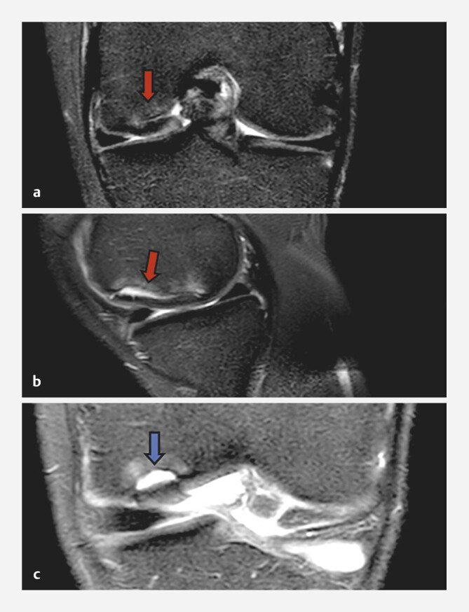
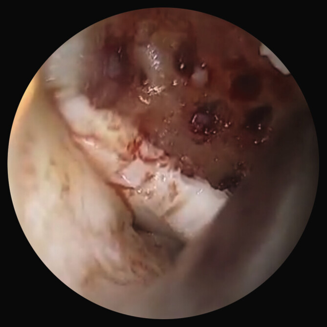
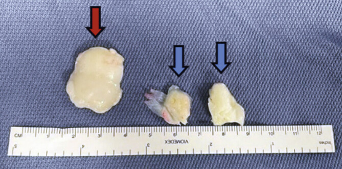
Methods: We describe a case of a patient suffering from OD with a bone lesion in the weight-bearing area of medial femoral condyle. Arthroscopy was performed and an osteochondral fragment from the medial femoral condyle was observed and two articular loose bodies were removed. After months, the patient returned with pain and a locked knee. magnetic resonance imaging (MRI) presented a new unstable chondral flap at the posterior border of the previous lesion. Surgery was performed again, and at open examination, the previous OD lesions were covered by regenerative tissue, with a lesion of 3 cm2 at the inferior medial part of the chondral flap. The peripheral margins were cleaned, and a subchondral crater was curetted. The subchondral lesion was debrided, and the flap was fixed with pins and a central bioresorbable screws.
Results: Revision surgery with fixation of the chondral flap using bioresorbable pins and screws led to satisfactory results.
Conclusion: Open revision surgery allowed us a more accurate assessment of the OD area to provide an effective fixation of the chondral flap and in this circumstance, this should have been done after seeing the first MRI.
背景:骨软骨炎(OD)是膝关节软骨最常见的病变之一。如果病变稳定,无松动体或有开放性髌骨,建议采用保守治疗。对于软骨病变不稳定或伴有松动体的无症状成人,建议以手术治疗为主:我们描述了一例股骨内侧髁负重区骨质病变的 OD 患者。我们在关节镜下观察到股骨内侧髁的骨软骨碎片,并取出了两个关节松动体。几个月后,患者因疼痛和膝关节锁定复诊。磁共振成像(MRI)显示,在之前病变的后缘有一个新的不稳定软骨瓣。再次进行手术,在开放性检查时,之前的外径病变被再生组织覆盖,软骨瓣下内侧有一个 3 平方厘米的病变。对周围边缘进行了清理,并刮除了软骨下凹坑。对软骨下病灶进行了清创,并用销钉和中心生物可吸收螺钉固定了软骨瓣:使用生物可吸收钉和螺钉固定软骨瓣的翻修手术取得了令人满意的效果:结论:开放式翻修手术让我们能够更准确地评估外径区域,从而有效固定软骨瓣,在这种情况下,应该在看到第一次核磁共振成像后再进行翻修手术。
期刊介绍:
Das Forum für Orthopädie und Unfallchirurgie aus einer Hand
Aktuelles aus Klinik, Wissenschaft und Forschung
Ein unabhängiges Peer-Review-Verfahren sichert Qualität, Relevanz und Plausibilität der Daten
Modernes Layout: Klare Gliederung, farbige Abbildungen, strukturierte Tabellen
Orthopädie und Unfallchirurgie aktuell: Berichte und Reportagen zu den wichtigsten Themen im Fach


 分享
分享
 求助内容:
求助内容: 应助结果提醒方式:
 扫码关注我们
