Does Using Indocyanine Green Fluorescence Imaging for Tumors Help in Determining the Safe Surgical Margin in Real-Time Navigation of Laparoscopic Hepatectomy? A Retrospective Study
Xinran Cai MD, PhD, Haijie Hong MD, PhD, Wei Pan MD, Jiangzhi Chen MD, PhD, Lei Jiang MD, Qiang Du MD, PhD, Ge Li MD, Shengzhe Lin MD, Yanling Chen MD, PhD
{"title":"Does Using Indocyanine Green Fluorescence Imaging for Tumors Help in Determining the Safe Surgical Margin in Real-Time Navigation of Laparoscopic Hepatectomy? A Retrospective Study","authors":"Xinran Cai MD, PhD, Haijie Hong MD, PhD, Wei Pan MD, Jiangzhi Chen MD, PhD, Lei Jiang MD, Qiang Du MD, PhD, Ge Li MD, Shengzhe Lin MD, Yanling Chen MD, PhD","doi":"10.1245/s10434-022-12893-3","DOIUrl":null,"url":null,"abstract":"<div><h3>Background</h3><p>This study aims to investigate whether indocyanine green (ICG) tumor imaging helps determine the safe surgical margin in laparoscopic hepatectomy.</p><h3>Patients and Methods</h3><p>Eighty-six patients with hepatic malignancies [including hepatocellular carcinoma (HCC) and colorectal liver metastasis (CRLM)] were included in this study. ICG-R15 testing was performed 5–7 days before surgery. Fluorescence staining of the tumor was detected by a fluorescent laparoscope, and the width of fluorescence band surrounding tumor was measured by an electronic vernier caliper.</p><h3>Results</h3><p>The positive rate of hepatic malignant lesions successfully stained by ICG fluorescence was 96.0% (95/99). HCC with better differentiation demonstrated non-rim fluorescence patterns, while cases with poor differentiation demonstrated rim patterns. CRLM uniformly demonstrated rim pattern. The width of fluorescence surrounding tumors was 0 in HCC with non-rim patterns. The minimum width of fluorescence surrounding tumors in poor differentiated HCC and CRLM were 2.4 ± 1.9 mm and 2.8 ± 2.5 mm, respectively, with no significant difference (<i>P</i> > 0.05). ICG fluorescence imaging revealed eight small lesions, which were not detected preoperatively in seven patients, of which five lesions were confirmed as malignancies by pathology.</p><h3>Conclusions</h3><p>Resection along the ICG fluorescence edge can supply a safe surgical margin only for CRLM, but not for HCC. Otherwise, ICG fluorescence tumor imaging can preliminarily determine the pathological type of hepatic malignancies and histological differentiation of HCC and help detect small lesions that cannot be detected preoperatively.</p></div>","PeriodicalId":8229,"journal":{"name":"Annals of Surgical Oncology","volume":"30 4","pages":"1981 - 1987"},"PeriodicalIF":3.5000,"publicationDate":"2022-12-09","publicationTypes":"Journal Article","fieldsOfStudy":null,"isOpenAccess":false,"openAccessPdf":"https://link.springer.com/content/pdf/10.1245/s10434-022-12893-3.pdf","citationCount":"2","resultStr":null,"platform":"Semanticscholar","paperid":null,"PeriodicalName":"Annals of Surgical Oncology","FirstCategoryId":"3","ListUrlMain":"https://link.springer.com/article/10.1245/s10434-022-12893-3","RegionNum":2,"RegionCategory":"医学","ArticlePicture":[],"TitleCN":null,"AbstractTextCN":null,"PMCID":null,"EPubDate":"","PubModel":"","JCR":"Q2","JCRName":"ONCOLOGY","Score":null,"Total":0}
引用次数: 2
Abstract
Background
This study aims to investigate whether indocyanine green (ICG) tumor imaging helps determine the safe surgical margin in laparoscopic hepatectomy.
Patients and Methods
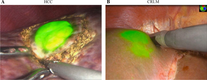
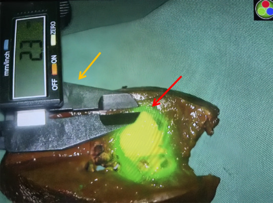
Eighty-six patients with hepatic malignancies [including hepatocellular carcinoma (HCC) and colorectal liver metastasis (CRLM)] were included in this study. ICG-R15 testing was performed 5–7 days before surgery. Fluorescence staining of the tumor was detected by a fluorescent laparoscope, and the width of fluorescence band surrounding tumor was measured by an electronic vernier caliper.
Results
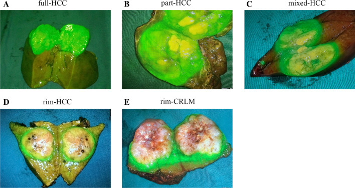
The positive rate of hepatic malignant lesions successfully stained by ICG fluorescence was 96.0% (95/99). HCC with better differentiation demonstrated non-rim fluorescence patterns, while cases with poor differentiation demonstrated rim patterns. CRLM uniformly demonstrated rim pattern. The width of fluorescence surrounding tumors was 0 in HCC with non-rim patterns. The minimum width of fluorescence surrounding tumors in poor differentiated HCC and CRLM were 2.4 ± 1.9 mm and 2.8 ± 2.5 mm, respectively, with no significant difference (P > 0.05). ICG fluorescence imaging revealed eight small lesions, which were not detected preoperatively in seven patients, of which five lesions were confirmed as malignancies by pathology.
Conclusions
Resection along the ICG fluorescence edge can supply a safe surgical margin only for CRLM, but not for HCC. Otherwise, ICG fluorescence tumor imaging can preliminarily determine the pathological type of hepatic malignancies and histological differentiation of HCC and help detect small lesions that cannot be detected preoperatively.
期刊介绍:
The Annals of Surgical Oncology is the official journal of The Society of Surgical Oncology and is published for the Society by Springer. The Annals publishes original and educational manuscripts about oncology for surgeons from all specialities in academic and community settings.


 分享
分享
 求助内容:
求助内容: 应助结果提醒方式:
 扫码关注我们
