Jaka Kragelj, Rupam Ghosh, Yiling Xiao, Rania Dumarieh, Dominique Lagasca, Sakshi Krishna, Kendra K Frederick
{"title":"Spatially resolved DNP-assisted NMR illuminates the conformational ensemble of α-synuclein in intact viable cells.","authors":"Jaka Kragelj, Rupam Ghosh, Yiling Xiao, Rania Dumarieh, Dominique Lagasca, Sakshi Krishna, Kendra K Frederick","doi":"10.1101/2023.10.24.563877","DOIUrl":null,"url":null,"abstract":"<p><p>The protein α-syn adopts a wide variety of conformations including an intrinsically disordered monomeric form and an α-helical rich membrane-associated form that is thought to play an important role in cellular membrane processes. However, despite the high affinity of α-syn for membranes, evidence that the α-helical form is adopted inside cells has been indirect. DNP-assisted solid state NMR on frozen cellular samples can report on protein conformations inside cells. Moreover, by controlling the distribution of the DNP polarization agent throughout the cellular biomass, such experiments can provide quantitative information upon the entire structural ensemble or provide information about spatially resolved sub-populations. Using DNP-assisted magic angle spinning (MAS) NMR we establish that purified α-syn in the membrane-associated and intrinsically disordered forms have distinguishable spectra. We then introduced isotopically labeled monomeric α-syn into cells. When the DNP polarization agent is dispersed homogenously throughout the cell, we found that a minority of the α-syn inside cells adopted a highly α-helical rich conformation. When the DNP polarization agent is peripherally localized, we found that the α-helical rich conformation predominates. Thus, we provide direct evidence that α-helix rich conformations of α-syn are adopted near the cellular periphery inside cells under physiological conditions. Moreover, we demonstrate how selectively altering the spatial distribution of the DNP polarization agent can be a powerful tool to observe spatially distinct structural ensembles. This approach paves the way for more nuanced investigations into the conformations that proteins adopt in different areas of the cell.</p>","PeriodicalId":72407,"journal":{"name":"bioRxiv : the preprint server for biology","volume":" ","pages":""},"PeriodicalIF":0.0000,"publicationDate":"2025-01-04","publicationTypes":"Journal Article","fieldsOfStudy":null,"isOpenAccess":false,"openAccessPdf":"https://www.ncbi.nlm.nih.gov/pmc/articles/PMC10634803/pdf/","citationCount":"0","resultStr":null,"platform":"Semanticscholar","paperid":null,"PeriodicalName":"bioRxiv : the preprint server for biology","FirstCategoryId":"1085","ListUrlMain":"https://doi.org/10.1101/2023.10.24.563877","RegionNum":0,"RegionCategory":null,"ArticlePicture":[],"TitleCN":null,"AbstractTextCN":null,"PMCID":null,"EPubDate":"","PubModel":"","JCR":"","JCRName":"","Score":null,"Total":0}
引用次数: 0
Abstract
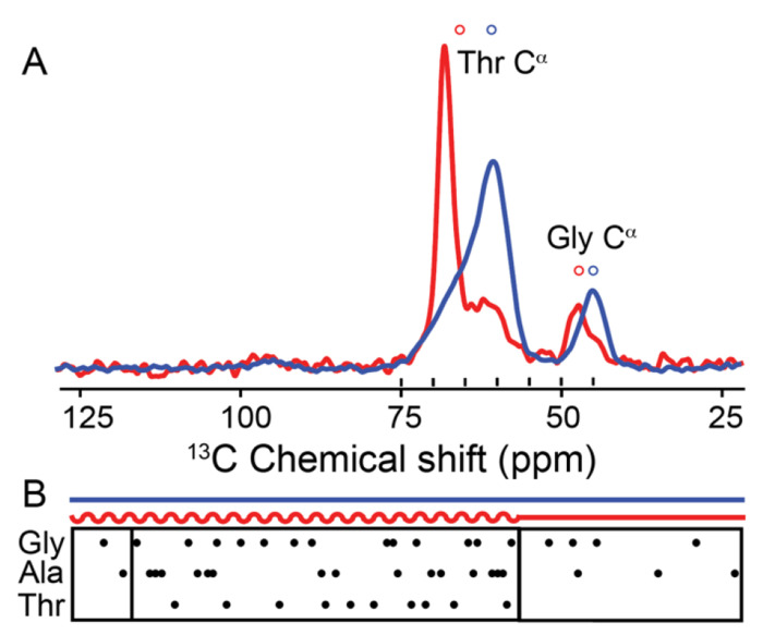
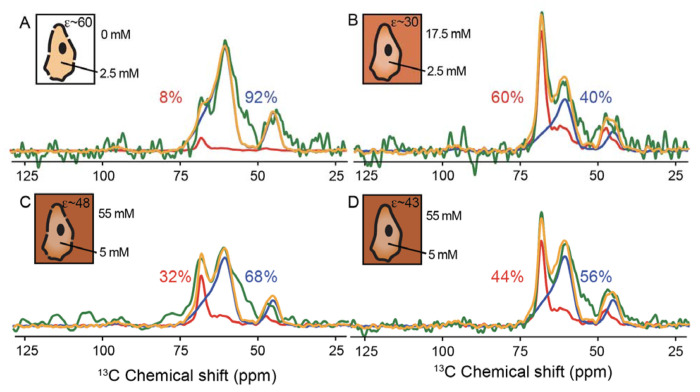
The protein α-syn adopts a wide variety of conformations including an intrinsically disordered monomeric form and an α-helical rich membrane-associated form that is thought to play an important role in cellular membrane processes. However, despite the high affinity of α-syn for membranes, evidence that the α-helical form is adopted inside cells has been indirect. DNP-assisted solid state NMR on frozen cellular samples can report on protein conformations inside cells. Moreover, by controlling the distribution of the DNP polarization agent throughout the cellular biomass, such experiments can provide quantitative information upon the entire structural ensemble or provide information about spatially resolved sub-populations. Using DNP-assisted magic angle spinning (MAS) NMR we establish that purified α-syn in the membrane-associated and intrinsically disordered forms have distinguishable spectra. We then introduced isotopically labeled monomeric α-syn into cells. When the DNP polarization agent is dispersed homogenously throughout the cell, we found that a minority of the α-syn inside cells adopted a highly α-helical rich conformation. When the DNP polarization agent is peripherally localized, we found that the α-helical rich conformation predominates. Thus, we provide direct evidence that α-helix rich conformations of α-syn are adopted near the cellular periphery inside cells under physiological conditions. Moreover, we demonstrate how selectively altering the spatial distribution of the DNP polarization agent can be a powerful tool to observe spatially distinct structural ensembles. This approach paves the way for more nuanced investigations into the conformations that proteins adopt in different areas of the cell.


 分享
分享

 求助内容:
求助内容: 应助结果提醒方式:
 扫码关注我们
