Magnetization transfer imaging of ovarian cancer: initial experiences of correlation with tissue cellularity and changes following neoadjuvant chemotherapy.
Surrin S Deen, Mary A McLean, Andrew B Gill, Robin A F Crawford, John Latimer, Peter Baldwin, Helena M Earl, Christine A Parkinson, Sarah Smith, Charlotte Hodgkin, Mercedes Jimenez-Linan, Cara R Brodie, Ilse Patterson, Helen C Addley, Susan J Freeman, Penelope M Moyle, Martin J Graves, Evis Sala, James D Brenton, Ferdia A Gallagher
{"title":"Magnetization transfer imaging of ovarian cancer: initial experiences of correlation with tissue cellularity and changes following neoadjuvant chemotherapy.","authors":"Surrin S Deen, Mary A McLean, Andrew B Gill, Robin A F Crawford, John Latimer, Peter Baldwin, Helena M Earl, Christine A Parkinson, Sarah Smith, Charlotte Hodgkin, Mercedes Jimenez-Linan, Cara R Brodie, Ilse Patterson, Helen C Addley, Susan J Freeman, Penelope M Moyle, Martin J Graves, Evis Sala, James D Brenton, Ferdia A Gallagher","doi":"10.1259/bjro.20210078","DOIUrl":null,"url":null,"abstract":"<p><strong>Objectives: </strong>To investigate the relationship between magnetization transfer (MT) imaging and tissue macromolecules in high-grade serous ovarian cancer (HGSOC) and whether MT ratio (MTR) changes following neoadjuvant chemotherapy (NACT).</p><p><strong>Methods: </strong>This was a prospective observational study. 12 HGSOC patients were imaged before treatment. MTR was compared to quantified tissue histology and immunohistochemistry. For a subset of patients (<i>n</i> = 5), MT imaging was repeated after NACT. The Shapiro-Wilk test was used to assess for normality of data and Spearman's rank-order or Pearson's correlation tests were then used to compare MTR with tissue quantifications. The Wilcoxon signed-rank test was used to assess for changes in MTR after treatment.</p><p><strong>Results: </strong>Treatment-naïve tumour MTR was 21.9 ± 3.1% (mean ± S.D.). MTR had a positive correlation with cellularity, rho = 0.56 (<i>p</i> < 0.05) and a negative correlation with tumour volume, ρ = -0.72 (<i>p</i> = 0.01). MTR did not correlate with the extracellular proteins, collagen IV or laminin (<i>p</i> = 0.40 and <i>p</i> = 0.90). For those patients imaged before and after NACT, an increase in MTR was observed in each case with mean MTR 20.6 ± 3.1% (median 21.1) pre-treatment and 25.6 ± 3.4% (median 26.5) post-treatment (<i>p</i> = 0.06).</p><p><strong>Conclusion: </strong>In treatment-naïve HGSOC, MTR is associated with cellularity, possibly reflecting intracellular macromolecular concentration. MT may also detect the HGSOC response to NACT, however larger studies are required to validate this finding.</p><p><strong>Advances in knowledge: </strong>MTR in HGSOC is influenced by cellularity. This may be applied to assess for cell changes following treatment.</p>","PeriodicalId":72419,"journal":{"name":"BJR open","volume":"4 1","pages":"20210078"},"PeriodicalIF":2.1000,"publicationDate":"2022-05-02","publicationTypes":"Journal Article","fieldsOfStudy":null,"isOpenAccess":false,"openAccessPdf":"https://www.ncbi.nlm.nih.gov/pmc/articles/PMC9459873/pdf/","citationCount":"0","resultStr":null,"platform":"Semanticscholar","paperid":null,"PeriodicalName":"BJR open","FirstCategoryId":"1085","ListUrlMain":"https://doi.org/10.1259/bjro.20210078","RegionNum":0,"RegionCategory":null,"ArticlePicture":[],"TitleCN":null,"AbstractTextCN":null,"PMCID":null,"EPubDate":"2022/1/1 0:00:00","PubModel":"eCollection","JCR":"","JCRName":"","Score":null,"Total":0}
引用次数: 0
Abstract
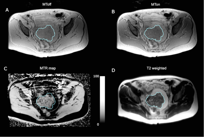
Objectives: To investigate the relationship between magnetization transfer (MT) imaging and tissue macromolecules in high-grade serous ovarian cancer (HGSOC) and whether MT ratio (MTR) changes following neoadjuvant chemotherapy (NACT).
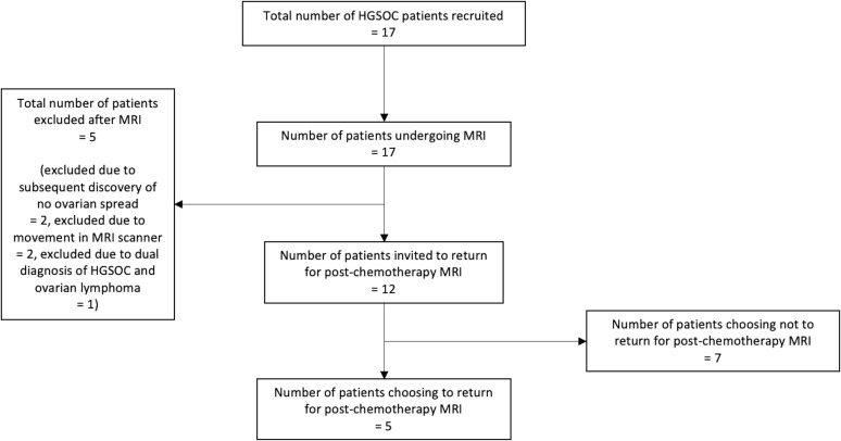
Methods: This was a prospective observational study. 12 HGSOC patients were imaged before treatment. MTR was compared to quantified tissue histology and immunohistochemistry. For a subset of patients (n = 5), MT imaging was repeated after NACT. The Shapiro-Wilk test was used to assess for normality of data and Spearman's rank-order or Pearson's correlation tests were then used to compare MTR with tissue quantifications. The Wilcoxon signed-rank test was used to assess for changes in MTR after treatment.
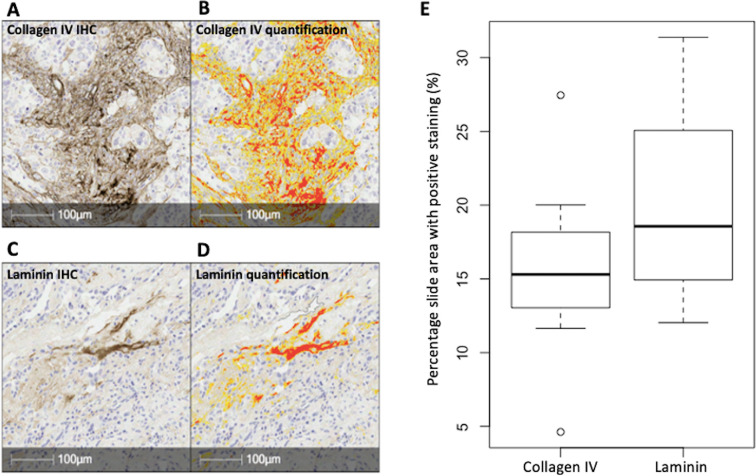
Results: Treatment-naïve tumour MTR was 21.9 ± 3.1% (mean ± S.D.). MTR had a positive correlation with cellularity, rho = 0.56 (p < 0.05) and a negative correlation with tumour volume, ρ = -0.72 (p = 0.01). MTR did not correlate with the extracellular proteins, collagen IV or laminin (p = 0.40 and p = 0.90). For those patients imaged before and after NACT, an increase in MTR was observed in each case with mean MTR 20.6 ± 3.1% (median 21.1) pre-treatment and 25.6 ± 3.4% (median 26.5) post-treatment (p = 0.06).
Conclusion: In treatment-naïve HGSOC, MTR is associated with cellularity, possibly reflecting intracellular macromolecular concentration. MT may also detect the HGSOC response to NACT, however larger studies are required to validate this finding.
Advances in knowledge: MTR in HGSOC is influenced by cellularity. This may be applied to assess for cell changes following treatment.


 分享
分享

 求助内容:
求助内容: 应助结果提醒方式:
 扫码关注我们
