Soonil Koun, Hye-Jin Park, Su-Min Jung, Jin Joo Cha, Dae Ryong Cha, Young Sun Kang
{"title":"Puromycin-induced kidney injury and subsequent regeneration in adult zebrafish.","authors":"Soonil Koun, Hye-Jin Park, Su-Min Jung, Jin Joo Cha, Dae Ryong Cha, Young Sun Kang","doi":"10.1080/19768354.2023.2203211","DOIUrl":null,"url":null,"abstract":"<p><p>Puromycin treatment can cause glomerular injury to the kidney, leading to proteinuria. However, the pathogenesis of acute kidney injury and subsequent regeneration after puromycin administration in animal models remain unclear. In this work, we examined the characteristics of kidney injury and subsequent regeneration following puromycin treatment in adult zebrafish. We intraperitoneally injected 100 μg of puromycin into zebrafish; sacrificed them at 1, 3, 5, 7, or 14 days post-injection (dpi); and examined the morphological, functional, and molecular changes in the kidney. Puromycin-treated zebrafish presented more rapid clearance of rhodamine dextran than control animals. Morphological changes were observed immediately after the puromycin injection (1-7 dpi) and had recovered by 14 dpi. The mRNA production of <i>lhx1a</i>, a renal progenitor marker, increased during recovery from kidney injury. Levels of NFκB, TNFα, Nampt, and p-ERK increased significantly during nephron injury and regeneration, and Sirt1, FOXO1, pax2, and wt1b showed an increasing tendency. However, TGF-β1 and smad5 production did not show any changes after puromycin treatment. This study provides evidence that puromycin-induced injury in adult zebrafish kidneys is a potential tool for evaluating the mechanism of nephron injury and subsequent regeneration.</p>","PeriodicalId":7804,"journal":{"name":"Animal Cells and Systems","volume":"27 1","pages":"112-119"},"PeriodicalIF":3.2000,"publicationDate":"2023-01-01","publicationTypes":"Journal Article","fieldsOfStudy":null,"isOpenAccess":false,"openAccessPdf":"https://ftp.ncbi.nlm.nih.gov/pub/pmc/oa_pdf/7c/17/TACS_27_2203211.PMC10120544.pdf","citationCount":"0","resultStr":null,"platform":"Semanticscholar","paperid":null,"PeriodicalName":"Animal Cells and Systems","FirstCategoryId":"99","ListUrlMain":"https://doi.org/10.1080/19768354.2023.2203211","RegionNum":2,"RegionCategory":"生物学","ArticlePicture":[],"TitleCN":null,"AbstractTextCN":null,"PMCID":null,"EPubDate":"","PubModel":"","JCR":"Q3","JCRName":"CELL BIOLOGY","Score":null,"Total":0}
引用次数: 0
Abstract
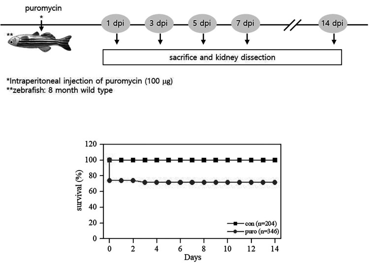
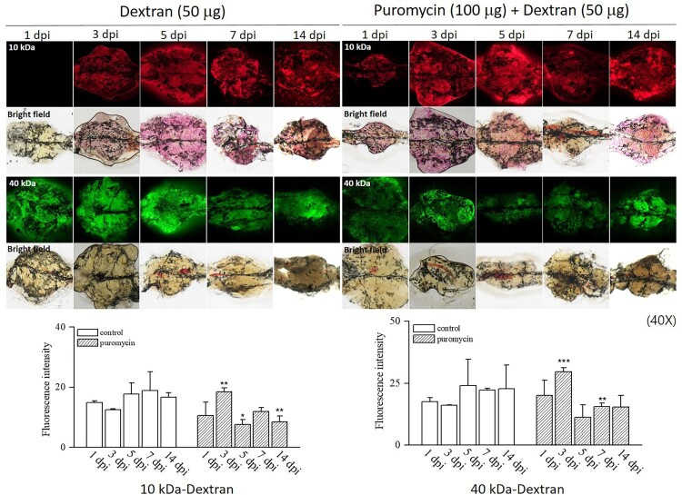
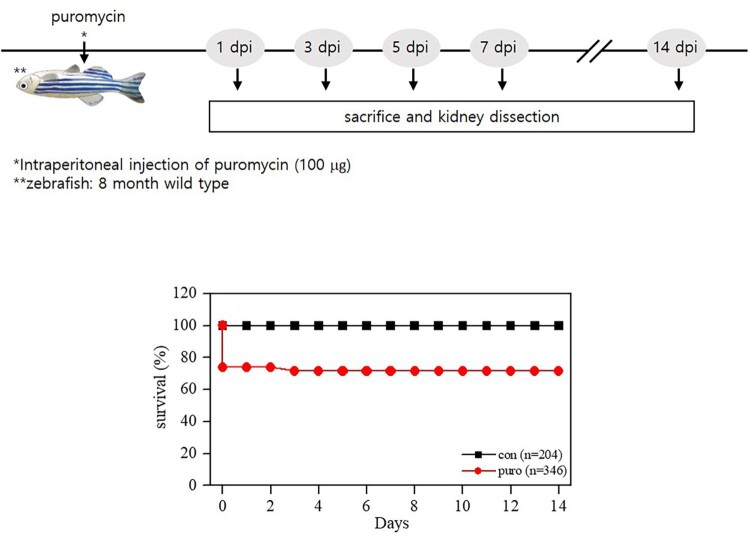
Puromycin treatment can cause glomerular injury to the kidney, leading to proteinuria. However, the pathogenesis of acute kidney injury and subsequent regeneration after puromycin administration in animal models remain unclear. In this work, we examined the characteristics of kidney injury and subsequent regeneration following puromycin treatment in adult zebrafish. We intraperitoneally injected 100 μg of puromycin into zebrafish; sacrificed them at 1, 3, 5, 7, or 14 days post-injection (dpi); and examined the morphological, functional, and molecular changes in the kidney. Puromycin-treated zebrafish presented more rapid clearance of rhodamine dextran than control animals. Morphological changes were observed immediately after the puromycin injection (1-7 dpi) and had recovered by 14 dpi. The mRNA production of lhx1a, a renal progenitor marker, increased during recovery from kidney injury. Levels of NFκB, TNFα, Nampt, and p-ERK increased significantly during nephron injury and regeneration, and Sirt1, FOXO1, pax2, and wt1b showed an increasing tendency. However, TGF-β1 and smad5 production did not show any changes after puromycin treatment. This study provides evidence that puromycin-induced injury in adult zebrafish kidneys is a potential tool for evaluating the mechanism of nephron injury and subsequent regeneration.
期刊介绍:
Animal Cells and Systems is the official journal of the Korean Society for Integrative Biology. This international, peer-reviewed journal publishes original papers that cover diverse aspects of biological sciences including Bioinformatics and Systems Biology, Developmental Biology, Evolution and Systematic Biology, Population Biology, & Animal Behaviour, Molecular and Cellular Biology, Neurobiology and Immunology, and Translational Medicine.


 分享
分享
 求助内容:
求助内容: 应助结果提醒方式:
 扫码关注我们
