{"title":"Design of a Machine Learning System to Predict the Thickness of a Melanoma Lesion in a Non-Invasive Way from Dermoscopic Images.","authors":"Ádám Szijártó, Ellák Somfai, András Lőrincz","doi":"10.4258/hir.2023.29.2.112","DOIUrl":null,"url":null,"abstract":"<p><strong>Objectives: </strong>Melanoma is the deadliest form of skin cancer, but it can be fully cured through early detection and treatment in 99% of cases. Our aim was to develop a non-invasive machine learning system that can predict the thickness of a melanoma lesion, which is a proxy for tumor progression, through dermoscopic images. This method can serve as a valuable tool in identifying urgent cases for treatment.</p><p><strong>Methods: </strong>A modern convolutional neural network architecture (EfficientNet) was used to construct a model capable of classifying dermoscopic images of melanoma lesions into three distinct categories based on thickness. We incorporated techniques to reduce the impact of an imbalanced training dataset, enhanced the generalization capacity of the model through image augmentation, and utilized five-fold cross-validation to produce more reliable metrics.</p><p><strong>Results: </strong>Our method achieved 71% balanced accuracy for three-way classification when trained on a small public dataset of 247 melanoma images. We also presented performance projections for larger training datasets.</p><p><strong>Conclusions: </strong>Our model represents a new state-of-the-art method for classifying melanoma thicknesses. Performance can be further optimized by expanding training datasets and utilizing model ensembles. We have shown that earlier claims of higher performance were mistaken due to data leakage during the evaluation process.</p>","PeriodicalId":12947,"journal":{"name":"Healthcare Informatics Research","volume":"29 2","pages":"112-119"},"PeriodicalIF":2.1000,"publicationDate":"2023-04-01","publicationTypes":"Journal Article","fieldsOfStudy":null,"isOpenAccess":false,"openAccessPdf":"https://ftp.ncbi.nlm.nih.gov/pub/pmc/oa_pdf/68/55/hir-2023-29-2-112.PMC10209725.pdf","citationCount":"2","resultStr":null,"platform":"Semanticscholar","paperid":null,"PeriodicalName":"Healthcare Informatics Research","FirstCategoryId":"1085","ListUrlMain":"https://doi.org/10.4258/hir.2023.29.2.112","RegionNum":0,"RegionCategory":null,"ArticlePicture":[],"TitleCN":null,"AbstractTextCN":null,"PMCID":null,"EPubDate":"","PubModel":"","JCR":"Q3","JCRName":"MEDICAL INFORMATICS","Score":null,"Total":0}
引用次数: 2
Abstract
Objectives: Melanoma is the deadliest form of skin cancer, but it can be fully cured through early detection and treatment in 99% of cases. Our aim was to develop a non-invasive machine learning system that can predict the thickness of a melanoma lesion, which is a proxy for tumor progression, through dermoscopic images. This method can serve as a valuable tool in identifying urgent cases for treatment.
Methods: A modern convolutional neural network architecture (EfficientNet) was used to construct a model capable of classifying dermoscopic images of melanoma lesions into three distinct categories based on thickness. We incorporated techniques to reduce the impact of an imbalanced training dataset, enhanced the generalization capacity of the model through image augmentation, and utilized five-fold cross-validation to produce more reliable metrics.
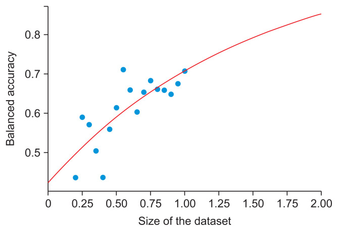
Results: Our method achieved 71% balanced accuracy for three-way classification when trained on a small public dataset of 247 melanoma images. We also presented performance projections for larger training datasets.
Conclusions: Our model represents a new state-of-the-art method for classifying melanoma thicknesses. Performance can be further optimized by expanding training datasets and utilizing model ensembles. We have shown that earlier claims of higher performance were mistaken due to data leakage during the evaluation process.


 分享
分享
 求助内容:
求助内容: 应助结果提醒方式:
 扫码关注我们
