{"title":"Automatic Method for Optic Disc Segmentation Using Deep Learning on Retinal Fundus Images.","authors":"Anindita Septiarini, Hamdani Hamdani, Emy Setyaningsih, Eko Junirianto, Fitri Utaminingrum","doi":"10.4258/hir.2023.29.2.145","DOIUrl":null,"url":null,"abstract":"<p><strong>Objectives: </strong>The optic disc is part of the retinal fundus image structure, which influences the extraction of glaucoma features. This study proposes a method that automatically segments the optic disc area in retinal fundus images using deep learning based on a convolutional neural network (CNN).</p><p><strong>Methods: </strong>This study used private and public datasets containing retinal fundus images. The private dataset consisted of 350 images, while the public dataset was the Retinal Fundus Glaucoma Challenge (REFUGE). The proposed method was based on a CNN with a single-shot multibox detector (MobileNetV2) to form images of the region-of-interest (ROI) using the original image resized into 640 × 640 input data. A pre-processing sequence was then implemented, including augmentation, resizing, and normalization. Furthermore, a U-Net model was applied for optic disc segmentation with 128 × 128 input data.</p><p><strong>Results: </strong>The proposed method was appropriately applied to the datasets used, as shown by the values of the F1-score, dice score, and intersection over union of 0.9880, 0.9852, and 0.9763 for the private dataset, respectively, and 0.9854, 0.9838 and 0.9712 for the REFUGE dataset.</p><p><strong>Conclusions: </strong>The optic disc area produced by the proposed method was similar to that identified by an ophthalmologist. Therefore, this method can be considered for implementing automatic segmentation of the optic disc area.</p>","PeriodicalId":12947,"journal":{"name":"Healthcare Informatics Research","volume":"29 2","pages":"145-151"},"PeriodicalIF":2.1000,"publicationDate":"2023-04-01","publicationTypes":"Journal Article","fieldsOfStudy":null,"isOpenAccess":false,"openAccessPdf":"https://ftp.ncbi.nlm.nih.gov/pub/pmc/oa_pdf/f1/52/hir-2023-29-2-145.PMC10209731.pdf","citationCount":"1","resultStr":null,"platform":"Semanticscholar","paperid":null,"PeriodicalName":"Healthcare Informatics Research","FirstCategoryId":"1085","ListUrlMain":"https://doi.org/10.4258/hir.2023.29.2.145","RegionNum":0,"RegionCategory":null,"ArticlePicture":[],"TitleCN":null,"AbstractTextCN":null,"PMCID":null,"EPubDate":"","PubModel":"","JCR":"Q3","JCRName":"MEDICAL INFORMATICS","Score":null,"Total":0}
引用次数: 1
Abstract
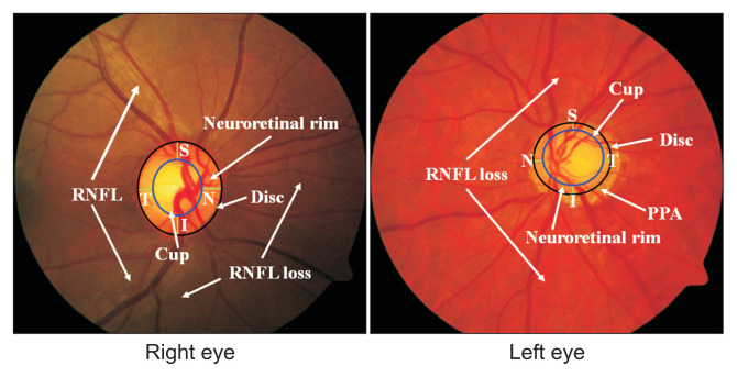
Objectives: The optic disc is part of the retinal fundus image structure, which influences the extraction of glaucoma features. This study proposes a method that automatically segments the optic disc area in retinal fundus images using deep learning based on a convolutional neural network (CNN).
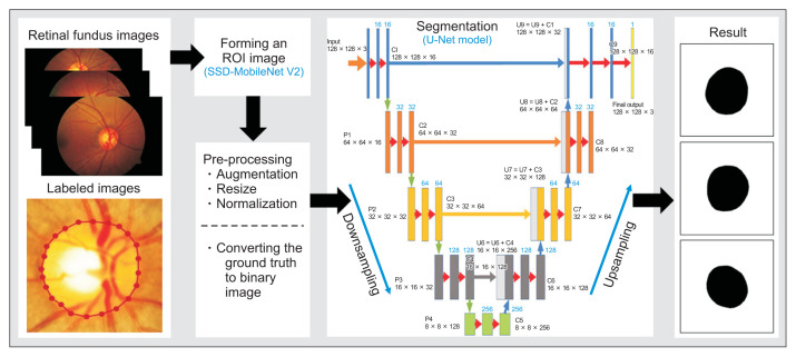
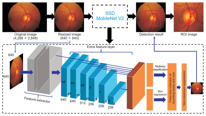
Methods: This study used private and public datasets containing retinal fundus images. The private dataset consisted of 350 images, while the public dataset was the Retinal Fundus Glaucoma Challenge (REFUGE). The proposed method was based on a CNN with a single-shot multibox detector (MobileNetV2) to form images of the region-of-interest (ROI) using the original image resized into 640 × 640 input data. A pre-processing sequence was then implemented, including augmentation, resizing, and normalization. Furthermore, a U-Net model was applied for optic disc segmentation with 128 × 128 input data.
Results: The proposed method was appropriately applied to the datasets used, as shown by the values of the F1-score, dice score, and intersection over union of 0.9880, 0.9852, and 0.9763 for the private dataset, respectively, and 0.9854, 0.9838 and 0.9712 for the REFUGE dataset.
Conclusions: The optic disc area produced by the proposed method was similar to that identified by an ophthalmologist. Therefore, this method can be considered for implementing automatic segmentation of the optic disc area.


 分享
分享
 求助内容:
求助内容: 应助结果提醒方式:
 扫码关注我们
