{"title":"Microcystic Meningioma - A Diagnostic Dilemma During Intraoperative Squash Smear Study.","authors":"Sukhpreet Kaur, Rahul Karode, Hanni V Gulwani","doi":"10.4103/joc.joc_28_22","DOIUrl":null,"url":null,"abstract":"<p><strong>Background: </strong>Meningiomas are generally slow-growing, benign, and non-infiltrating in nature. They are usually easy to diagnose cytologically if they are of the meningothelial type; however, they may cause diagnostic challenges when they manifest as unusual morphological variants, like the microcystic type. Because of the rarity of microcystic meningioma (MM), information on its cytological features is rarely available in the literature.</p><p><strong>Objectives: </strong>The goal of this study is to review the cytological features of MM in crush preparations prepared at the time of intra-op consultation and to identify the more common features which are helpful in rendering a correct diagnosis.</p><p><strong>Material and methods: </strong>Cytological features of five cases of MM were reviewed and noted from the records.</p><p><strong>Results: </strong>There were five patients of MM with a male: female ratio of 1.5:1 and a mean age of 52 years. All tumors were supratentorial and dura-based. Magnetic resonance imaging (MRI) showed low signal intensity on T1 and high signal intensity on T2 weighted images in four cases. Cytosmears were moderate-to-highly cellular. There were variable-sized cystic spaces within the meningothelial cell clusters. In four cases, nuclear pleomorphism was frequently observed. Nuclear pseudoinclusions, atypical mitoses, vascular proliferation, and necrosis were absent in all cases. Whorling and psammoma bodies were seen in only one case.</p><p><strong>Conclusion: </strong>Cytological features identified would be helpful in the diagnosis of microcystic meningiomas, especially in unusual radiological findings. Their unusual cytological features might lead to problems in differential diagnosis from other intracranial tumors, including glioblastoma, metastatic tumor, etc.</p>","PeriodicalId":50217,"journal":{"name":"Journal of Cytology","volume":"40 1","pages":"19-23"},"PeriodicalIF":1.0000,"publicationDate":"2023-01-01","publicationTypes":"Journal Article","fieldsOfStudy":null,"isOpenAccess":false,"openAccessPdf":"https://www.ncbi.nlm.nih.gov/pmc/articles/PMC10167828/pdf/","citationCount":"1","resultStr":null,"platform":"Semanticscholar","paperid":null,"PeriodicalName":"Journal of Cytology","FirstCategoryId":"3","ListUrlMain":"https://doi.org/10.4103/joc.joc_28_22","RegionNum":4,"RegionCategory":"医学","ArticlePicture":[],"TitleCN":null,"AbstractTextCN":null,"PMCID":null,"EPubDate":"2023/2/15 0:00:00","PubModel":"Epub","JCR":"Q4","JCRName":"MEDICAL LABORATORY TECHNOLOGY","Score":null,"Total":0}
引用次数: 1
Abstract
Background: Meningiomas are generally slow-growing, benign, and non-infiltrating in nature. They are usually easy to diagnose cytologically if they are of the meningothelial type; however, they may cause diagnostic challenges when they manifest as unusual morphological variants, like the microcystic type. Because of the rarity of microcystic meningioma (MM), information on its cytological features is rarely available in the literature.
Objectives: The goal of this study is to review the cytological features of MM in crush preparations prepared at the time of intra-op consultation and to identify the more common features which are helpful in rendering a correct diagnosis.
Material and methods: Cytological features of five cases of MM were reviewed and noted from the records.
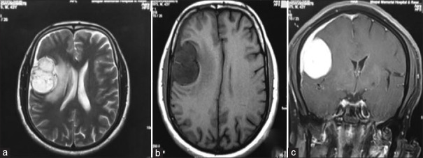
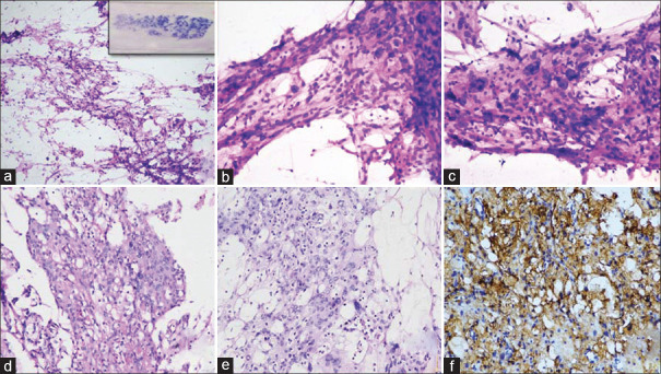
Results: There were five patients of MM with a male: female ratio of 1.5:1 and a mean age of 52 years. All tumors were supratentorial and dura-based. Magnetic resonance imaging (MRI) showed low signal intensity on T1 and high signal intensity on T2 weighted images in four cases. Cytosmears were moderate-to-highly cellular. There were variable-sized cystic spaces within the meningothelial cell clusters. In four cases, nuclear pleomorphism was frequently observed. Nuclear pseudoinclusions, atypical mitoses, vascular proliferation, and necrosis were absent in all cases. Whorling and psammoma bodies were seen in only one case.
Conclusion: Cytological features identified would be helpful in the diagnosis of microcystic meningiomas, especially in unusual radiological findings. Their unusual cytological features might lead to problems in differential diagnosis from other intracranial tumors, including glioblastoma, metastatic tumor, etc.
期刊介绍:
The Journal of Cytology is the official Quarterly publication of the Indian Academy of Cytologists. It is in the 25th year of publication in the year 2008. The journal covers all aspects of diagnostic cytology, including fine needle aspiration cytology, gynecological and non-gynecological cytology. Articles on ancillary techniques, like cytochemistry, immunocytochemistry, electron microscopy, molecular cytopathology, as applied to cytological material are also welcome. The journal gives preference to clinically oriented studies over experimental and animal studies. The Journal would publish peer-reviewed original research papers, case reports, systematic reviews, meta-analysis, and debates.


 分享
分享
 求助内容:
求助内容: 应助结果提醒方式:
 扫码关注我们
