Elizabeth I. Harper, Tyvette S. Hilliard, Emma F. Sheedy, Preston Carey, Paul Wilkinson, Michael D. Siroky, Jing Yang, Elizabeth Agadi, Annemarie K. Leonard, Ethan Low, Yueying Liu, Arya Biragyn, Christina M. Annunziata, Mary Sharon Stack
{"title":"Another wrinkle with age: Aged collagen and intra-peritoneal metastasis of ovarian cancer","authors":"Elizabeth I. Harper, Tyvette S. Hilliard, Emma F. Sheedy, Preston Carey, Paul Wilkinson, Michael D. Siroky, Jing Yang, Elizabeth Agadi, Annemarie K. Leonard, Ethan Low, Yueying Liu, Arya Biragyn, Christina M. Annunziata, Mary Sharon Stack","doi":"10.1002/aac2.12049","DOIUrl":null,"url":null,"abstract":"<div>\n \n \n <section>\n \n <h3> Background</h3>\n \n <p>Age is the most significant risk factor for ovarian cancer (OvCa), the deadliest gynecologic malignancy. Metastasizing OvCa cells adhere to the omentum, a peritoneal structure rich in collagen, adipocytes, and immune cells. Ultrastructural changes in the omentum and the omental collagen matrix with aging have not been evaluated.</p>\n </section>\n \n <section>\n \n <h3> Aim</h3>\n \n <p>The aim of this study was to test the hypothesis that age-related changes in collagen in the ovarian tumor microenvironment promote OvCa metastatic success in the aged host.</p>\n </section>\n \n <section>\n \n <h3> Methods/Results</h3>\n \n <p>Young (3–6 months) and aged mice (20–23 months) were used to study the role of aging in metastatic success. Intra-peritoneal (IP) injection of ID8<i>Trp53<sup>–/–</sup></i> OvCa cells showed enhanced IP dissemination in aged versus young mice. In vitro assays using purified collagen demonstrated reduced collagenolysis of aged fibers, as visualized using scanning electron microscopy (SEM) and quantified with a hydroxyproline release assay. Omental tumors in young and aged mice showed similar collagen deposition; however enhanced intra-tumoral collagen remodeling was seen in aged mice probed with a biotinylated collagen hybridizing peptide (CHP). In contrast, second harmonic generation (SHG) microscopy showed significant differences in collagen fiber structure and organization in omental tissue, and SEM demonstrated enhanced omental fenestration in aged omenta. Combined SHG and Alexa Fluor-CHP microscopy in vivo demonstrated that peri-tumoral collagen was remodeled more extensively in young mice. This collagen population represents truly aged host collagen, in contrast to intra-tumoral collagen that is newly synthesized, likely by cancer-associated fibroblasts.</p>\n </section>\n \n <section>\n \n <h3> Conclusions</h3>\n \n <p>Our results demonstrate that tumors in an aged host can grow with minimal collagen remodeling, while tumors in the young host must remodel peri-tumoral collagen to enable effective proliferation, providing a mechanism whereby age-induced ultrastructural changes in collagen and collagen-rich omenta establish a permissive premetastatic niche contributing to enhanced OvCa metastatic success in the aged host.</p>\n </section>\n </div>","PeriodicalId":72128,"journal":{"name":"Aging and cancer","volume":"3 2","pages":"116-129"},"PeriodicalIF":0.0000,"publicationDate":"2022-04-19","publicationTypes":"Journal Article","fieldsOfStudy":null,"isOpenAccess":false,"openAccessPdf":"https://www.ncbi.nlm.nih.gov/pmc/articles/PMC9518742/pdf/nihms-1793530.pdf","citationCount":"2","resultStr":null,"platform":"Semanticscholar","paperid":null,"PeriodicalName":"Aging and cancer","FirstCategoryId":"1085","ListUrlMain":"https://onlinelibrary.wiley.com/doi/10.1002/aac2.12049","RegionNum":0,"RegionCategory":null,"ArticlePicture":[],"TitleCN":null,"AbstractTextCN":null,"PMCID":null,"EPubDate":"","PubModel":"","JCR":"","JCRName":"","Score":null,"Total":0}
引用次数: 2
Abstract
Background
Age is the most significant risk factor for ovarian cancer (OvCa), the deadliest gynecologic malignancy. Metastasizing OvCa cells adhere to the omentum, a peritoneal structure rich in collagen, adipocytes, and immune cells. Ultrastructural changes in the omentum and the omental collagen matrix with aging have not been evaluated.
Aim
The aim of this study was to test the hypothesis that age-related changes in collagen in the ovarian tumor microenvironment promote OvCa metastatic success in the aged host.
Methods/Results
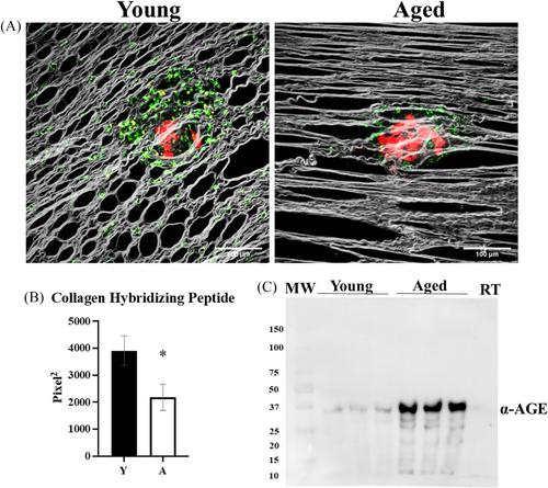
Young (3–6 months) and aged mice (20–23 months) were used to study the role of aging in metastatic success. Intra-peritoneal (IP) injection of ID8Trp53–/– OvCa cells showed enhanced IP dissemination in aged versus young mice. In vitro assays using purified collagen demonstrated reduced collagenolysis of aged fibers, as visualized using scanning electron microscopy (SEM) and quantified with a hydroxyproline release assay. Omental tumors in young and aged mice showed similar collagen deposition; however enhanced intra-tumoral collagen remodeling was seen in aged mice probed with a biotinylated collagen hybridizing peptide (CHP). In contrast, second harmonic generation (SHG) microscopy showed significant differences in collagen fiber structure and organization in omental tissue, and SEM demonstrated enhanced omental fenestration in aged omenta. Combined SHG and Alexa Fluor-CHP microscopy in vivo demonstrated that peri-tumoral collagen was remodeled more extensively in young mice. This collagen population represents truly aged host collagen, in contrast to intra-tumoral collagen that is newly synthesized, likely by cancer-associated fibroblasts.
Conclusions
Our results demonstrate that tumors in an aged host can grow with minimal collagen remodeling, while tumors in the young host must remodel peri-tumoral collagen to enable effective proliferation, providing a mechanism whereby age-induced ultrastructural changes in collagen and collagen-rich omenta establish a permissive premetastatic niche contributing to enhanced OvCa metastatic success in the aged host.


 分享
分享

 求助内容:
求助内容: 应助结果提醒方式:
 扫码关注我们
