Nan Zhang, Ruixing Liu, Xiaowu Liu, Songlin Hou, Runan Dou, Xingchen Geng, Yan Li, Jingguo Li, Lei Zhu, Zhanrong Li
{"title":"Personalized 3D-printed amniotic fornical ring for ocular surface reconstruction.","authors":"Nan Zhang, Ruixing Liu, Xiaowu Liu, Songlin Hou, Runan Dou, Xingchen Geng, Yan Li, Jingguo Li, Lei Zhu, Zhanrong Li","doi":"10.18063/ijb.713","DOIUrl":null,"url":null,"abstract":"<p><p>In the present work, we used three-dimensional (3D) printing technology to make a polylactic acid (PLA) amniotic fornical ring (AFR) for ocular surface reconstruction. This work is a retrospective and interventional case series of patients with ocular surface diseases who underwent either personalized 3D-printed AFR-assisted amniotic membrane transplantation (AMT) or sutured AMT (SAMT). Patient epidemiology, treatment, operative duration, epithelial healing time, retention time, vision changes, morbidity, and costs were analyzed. Thirty-one patients (40 eyes) and 19 patients (22 eyes) were enrolled in the 3D-printed AFR group and the SAMT group, respectively. The clinical indications of AFR and SAMT were similar, such as corneal and/or conjunctival epithelial defects due to chemical burns, thermal burns, Stevens-Johnson syndrome (SJS), or toxic epidermal necrolysis (TEN). The mean dissolution time was 15 ± 11 days in the AFR group, compared with 14 ± 7 days in the SAMT group. The percentage of healed corneal area was 90.91% (66.10%-100.00%) for AFR and 93.67% (60.23%-100.00%) for SAMT. The median time for corneal epithelial healing was 14 (7-75) days in the AFR group and 30 (14-55) days in the suture AMT group. There were no significant differences in the initial visual acuity, final visual acuity, or improvement in visual acuity between the two groups. The operation duration in the AFR group was significantly shorter than that in the SAMT group. Regarding the cost analysis, the average cost per eye in the AFR group was significantly lower than that in the SAMT group. Furthermore, 3D-printed and sterile AFR showed no obvious side effects on the eyes. Our results suggested that 3D-printed PLA scaffolds could be used as an AFR device for ocular surface disease. In addition, personalized 3D-printed AFR is superior to conventional AMT in operation duration and cost effectiveness, thereby reducing the financial burden on our health care system.</p>","PeriodicalId":48522,"journal":{"name":"International Journal of Bioprinting","volume":"9 3","pages":"713"},"PeriodicalIF":6.0000,"publicationDate":"2023-01-01","publicationTypes":"Journal Article","fieldsOfStudy":null,"isOpenAccess":false,"openAccessPdf":"https://ftp.ncbi.nlm.nih.gov/pub/pmc/oa_pdf/3b/2c/IJB-9-3-713.PMC10236349.pdf","citationCount":"1","resultStr":null,"platform":"Semanticscholar","paperid":null,"PeriodicalName":"International Journal of Bioprinting","FirstCategoryId":"5","ListUrlMain":"https://doi.org/10.18063/ijb.713","RegionNum":3,"RegionCategory":"医学","ArticlePicture":[],"TitleCN":null,"AbstractTextCN":null,"PMCID":null,"EPubDate":"","PubModel":"","JCR":"Q1","JCRName":"ENGINEERING, BIOMEDICAL","Score":null,"Total":0}
引用次数: 1
Abstract
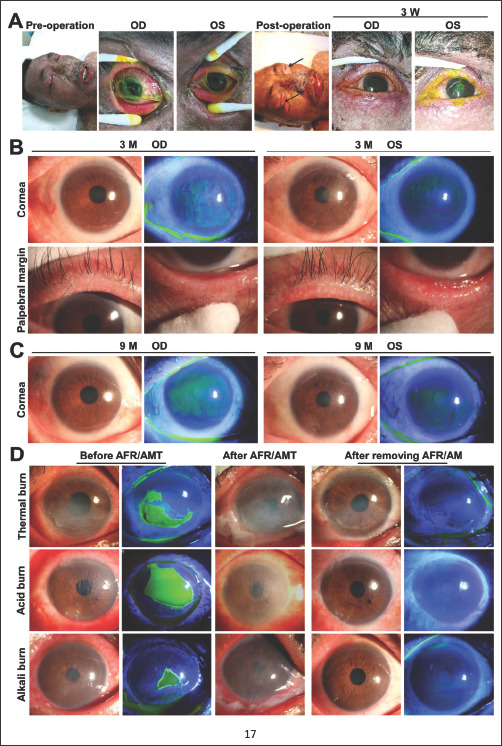
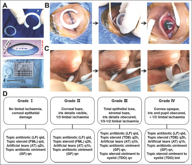
In the present work, we used three-dimensional (3D) printing technology to make a polylactic acid (PLA) amniotic fornical ring (AFR) for ocular surface reconstruction. This work is a retrospective and interventional case series of patients with ocular surface diseases who underwent either personalized 3D-printed AFR-assisted amniotic membrane transplantation (AMT) or sutured AMT (SAMT). Patient epidemiology, treatment, operative duration, epithelial healing time, retention time, vision changes, morbidity, and costs were analyzed. Thirty-one patients (40 eyes) and 19 patients (22 eyes) were enrolled in the 3D-printed AFR group and the SAMT group, respectively. The clinical indications of AFR and SAMT were similar, such as corneal and/or conjunctival epithelial defects due to chemical burns, thermal burns, Stevens-Johnson syndrome (SJS), or toxic epidermal necrolysis (TEN). The mean dissolution time was 15 ± 11 days in the AFR group, compared with 14 ± 7 days in the SAMT group. The percentage of healed corneal area was 90.91% (66.10%-100.00%) for AFR and 93.67% (60.23%-100.00%) for SAMT. The median time for corneal epithelial healing was 14 (7-75) days in the AFR group and 30 (14-55) days in the suture AMT group. There were no significant differences in the initial visual acuity, final visual acuity, or improvement in visual acuity between the two groups. The operation duration in the AFR group was significantly shorter than that in the SAMT group. Regarding the cost analysis, the average cost per eye in the AFR group was significantly lower than that in the SAMT group. Furthermore, 3D-printed and sterile AFR showed no obvious side effects on the eyes. Our results suggested that 3D-printed PLA scaffolds could be used as an AFR device for ocular surface disease. In addition, personalized 3D-printed AFR is superior to conventional AMT in operation duration and cost effectiveness, thereby reducing the financial burden on our health care system.
期刊介绍:
The International Journal of Bioprinting is a globally recognized publication that focuses on the advancements, scientific discoveries, and practical implementations of Bioprinting. Bioprinting, in simple terms, involves the utilization of 3D printing technology and materials that contain living cells or biological components to fabricate tissues or other biotechnological products. Our journal encompasses interdisciplinary research that spans across technology, science, and clinical applications within the expansive realm of Bioprinting.


 分享
分享
 求助内容:
求助内容: 应助结果提醒方式:
 扫码关注我们
