Margaux Verdier , Jeremy Deverdun , Nicolas Menjot de Champfleur , Hugues Duffau , Philippe Lam , Thomas Dos Santos , Thomas Troalen , Bénédicte Maréchal , Till Huelnhagen , Emmanuelle Le Bars
{"title":"Evaluation of a nnU-Net type automated clinical volumetric tumor segmentation tool for diffuse low-grade glioma follow-up","authors":"Margaux Verdier , Jeremy Deverdun , Nicolas Menjot de Champfleur , Hugues Duffau , Philippe Lam , Thomas Dos Santos , Thomas Troalen , Bénédicte Maréchal , Till Huelnhagen , Emmanuelle Le Bars","doi":"10.1016/j.neurad.2023.05.008","DOIUrl":null,"url":null,"abstract":"<div><h3>Background and purpose</h3><p><span>Diffuse low-grade gliomas (DLGG) are characterized by a slow and continuous growth and always evolve towards an aggressive grade. Accurate prediction of the malignant transformation<span> is essential as it requires immediate therapeutic intervention. One of its most precise predictors is the velocity of diameter expansion (VDE). Currently, the VDE is estimated either by </span></span>linear measurements<span> or by manual delineation of the DLGG on T2 FLAIR acquisitions. However, because of the DLGG's infiltrative nature and its blurred contours, manual measures are challenging and variable, even for experts. Therefore we propose an automated segmentation algorithm using a 2D nnU-Net, to 1) gain time and 2) standardize VDE assessment.</span></p></div><div><h3>Materials and Methods</h3><p>The 2D nnU-Net was trained on 318 acquisitions (T2 FLAIR & 3DT1 longitudinal follow-up of 30 patients, including pre- & post-surgery acquisitions, different scanners, vendors, imaging parameters…). Automated vs. manual segmentation performance was evaluated on 167 acquisitions, and its clinical interest was validated by quantifying the amount of manual correction required after automated segmentation of 98 novel acquisitions.</p></div><div><h3>Results</h3><p>Automated segmentation showed a good performance with a mean Dice Similarity Coefficient (DSC) of 0.82±0.13 with manual segmentation and a substantial concordance between VDE calculations. Major manual corrections (i.e., DSC<0.7) were necessary only in 3/98 cases and 81% of the cases had a DSC>0.9.</p></div><div><h3>Conclusion</h3><p>The proposed automated segmentation algorithm can successfully segment DLGG on highly variable MRI data. Although manual corrections are sometimes necessary, it provides a reliable, standardized and time-winning support for VDE extraction to asses DLGG growth.</p></div>","PeriodicalId":50115,"journal":{"name":"Journal of Neuroradiology","volume":"51 1","pages":"Pages 16-23"},"PeriodicalIF":3.3000,"publicationDate":"2024-02-01","publicationTypes":"Journal Article","fieldsOfStudy":null,"isOpenAccess":false,"openAccessPdf":"","citationCount":"0","resultStr":null,"platform":"Semanticscholar","paperid":null,"PeriodicalName":"Journal of Neuroradiology","FirstCategoryId":"3","ListUrlMain":"https://www.sciencedirect.com/science/article/pii/S0150986123002134","RegionNum":3,"RegionCategory":"医学","ArticlePicture":[],"TitleCN":null,"AbstractTextCN":null,"PMCID":null,"EPubDate":"","PubModel":"","JCR":"Q2","JCRName":"CLINICAL NEUROLOGY","Score":null,"Total":0}
引用次数: 0
Abstract
Background and purpose
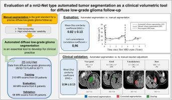
Diffuse low-grade gliomas (DLGG) are characterized by a slow and continuous growth and always evolve towards an aggressive grade. Accurate prediction of the malignant transformation is essential as it requires immediate therapeutic intervention. One of its most precise predictors is the velocity of diameter expansion (VDE). Currently, the VDE is estimated either by linear measurements or by manual delineation of the DLGG on T2 FLAIR acquisitions. However, because of the DLGG's infiltrative nature and its blurred contours, manual measures are challenging and variable, even for experts. Therefore we propose an automated segmentation algorithm using a 2D nnU-Net, to 1) gain time and 2) standardize VDE assessment.
Materials and Methods
The 2D nnU-Net was trained on 318 acquisitions (T2 FLAIR & 3DT1 longitudinal follow-up of 30 patients, including pre- & post-surgery acquisitions, different scanners, vendors, imaging parameters…). Automated vs. manual segmentation performance was evaluated on 167 acquisitions, and its clinical interest was validated by quantifying the amount of manual correction required after automated segmentation of 98 novel acquisitions.
Results
Automated segmentation showed a good performance with a mean Dice Similarity Coefficient (DSC) of 0.82±0.13 with manual segmentation and a substantial concordance between VDE calculations. Major manual corrections (i.e., DSC<0.7) were necessary only in 3/98 cases and 81% of the cases had a DSC>0.9.
Conclusion
The proposed automated segmentation algorithm can successfully segment DLGG on highly variable MRI data. Although manual corrections are sometimes necessary, it provides a reliable, standardized and time-winning support for VDE extraction to asses DLGG growth.
期刊介绍:
The Journal of Neuroradiology is a peer-reviewed journal, publishing worldwide clinical and basic research in the field of diagnostic and Interventional neuroradiology, translational and molecular neuroimaging, and artificial intelligence in neuroradiology.
The Journal of Neuroradiology considers for publication articles, reviews, technical notes and letters to the editors (correspondence section), provided that the methodology and scientific content are of high quality, and that the results will have substantial clinical impact and/or physiological importance.


 分享
分享
 求助内容:
求助内容: 应助结果提醒方式:
 扫码关注我们
