Jee Won Chai, Joo-Ho Lee, Dong Hyun Kim, Jina Park, So-Hee Oh, Su-Mi Shin
{"title":"Effect of Patient's Positioning on the Grade of Tendinosis and Visible Range of Infraspinatus Tendon on Ultrasound.","authors":"Jee Won Chai, Joo-Ho Lee, Dong Hyun Kim, Jina Park, So-Hee Oh, Su-Mi Shin","doi":"10.3348/jksr.2022.0137","DOIUrl":null,"url":null,"abstract":"<p><strong>Purpose: </strong>To investigate the effect of patient positioning on tendinosis grade, visible range, and infraspinatus tendon (IST) thickness, and to determine the feasibility of internal rotation (IR) position to assess IST on ultrasound (US).</p><p><strong>Materials and methods: </strong>This study included 52 shoulders of 48 subjects who were evaluated for IST in three different positions: neutral position (N), IR, and position with the ipsilateral hand on the contralateral shoulder (HC). Two radiologists retrospectively graded IST tendinosis from grade 0 to grade 3 and the visible range from grade 1 to grade 4. The thickness of the IST was measured by another radiologist with a short-axis view. A generalized estimating equation was used for statistical analysis.</p><p><strong>Results: </strong>The tendinosis grades were higher in the HC position than in the IR position, with a cumulative odds ratio of 2.087 (0.004, 95% confidence interval [CI]: 1.268-3.433). The tendinosis grades in the HC position (<i>p</i> = 0.370) and IR position (<i>p</i> = 0.146) were not significantly different from those in the N position. The overall difference in IST thickness was significant (<i>p</i> < 0.001), but the visible range (<i>p</i> = 0.530) was not significantly different according to position.</p><p><strong>Conclusion: </strong>Patient positioning significantly affected the grade of tendinosis and thickness but not the visible range of the IST. The IR position is a feasible position for assessing the IST on US.</p>","PeriodicalId":17455,"journal":{"name":"Journal of the Korean Society of Radiology","volume":"84 3","pages":"627-637"},"PeriodicalIF":0.0000,"publicationDate":"2023-05-01","publicationTypes":"Journal Article","fieldsOfStudy":null,"isOpenAccess":false,"openAccessPdf":"https://ftp.ncbi.nlm.nih.gov/pub/pmc/oa_pdf/14/d9/jksr-84-627.PMC10265234.pdf","citationCount":"0","resultStr":null,"platform":"Semanticscholar","paperid":null,"PeriodicalName":"Journal of the Korean Society of Radiology","FirstCategoryId":"1085","ListUrlMain":"https://doi.org/10.3348/jksr.2022.0137","RegionNum":0,"RegionCategory":null,"ArticlePicture":[],"TitleCN":null,"AbstractTextCN":null,"PMCID":null,"EPubDate":"","PubModel":"","JCR":"Q4","JCRName":"Medicine","Score":null,"Total":0}
引用次数: 0
Abstract
Purpose: To investigate the effect of patient positioning on tendinosis grade, visible range, and infraspinatus tendon (IST) thickness, and to determine the feasibility of internal rotation (IR) position to assess IST on ultrasound (US).
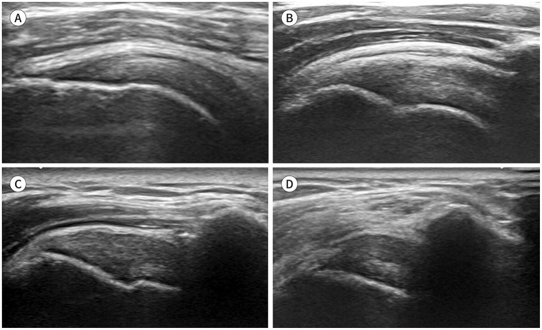
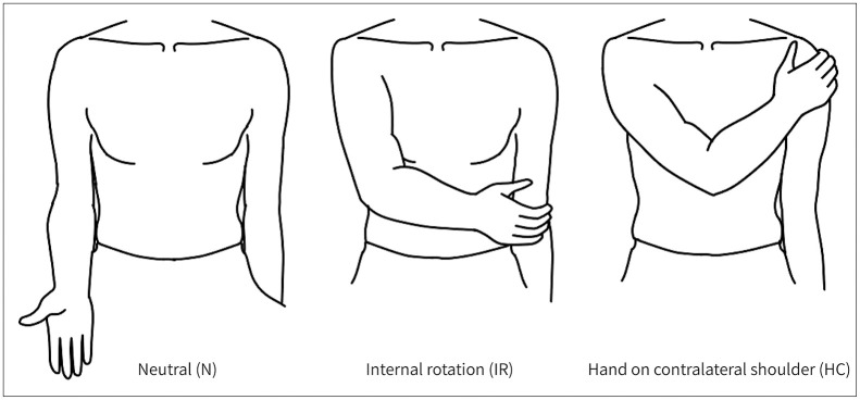
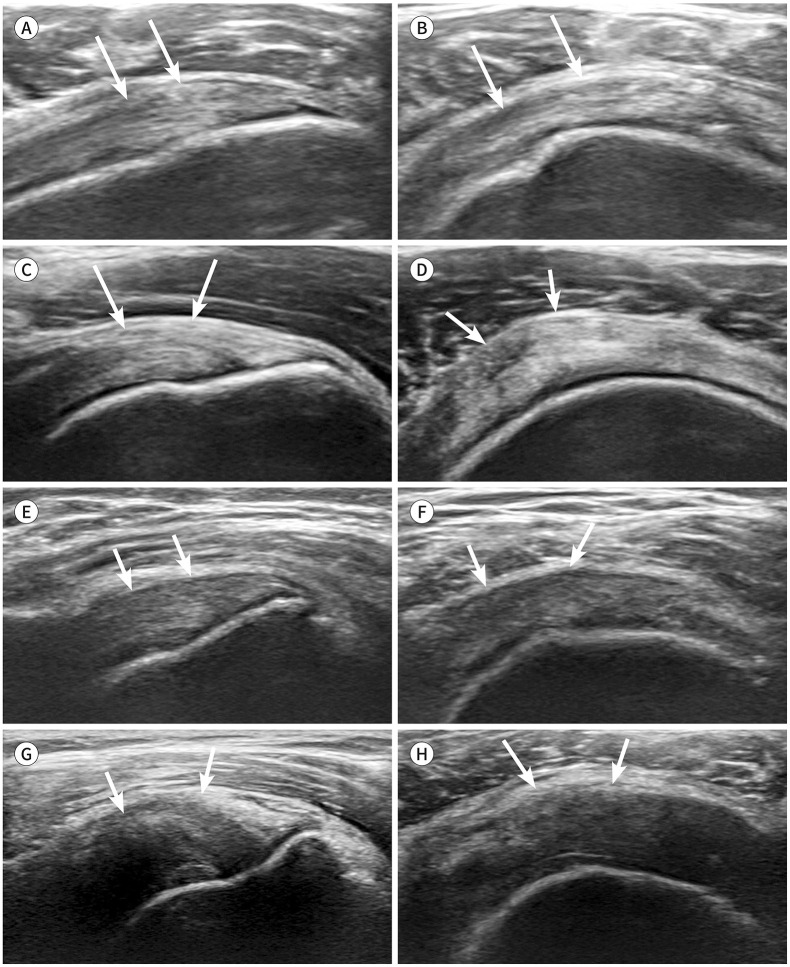
Materials and methods: This study included 52 shoulders of 48 subjects who were evaluated for IST in three different positions: neutral position (N), IR, and position with the ipsilateral hand on the contralateral shoulder (HC). Two radiologists retrospectively graded IST tendinosis from grade 0 to grade 3 and the visible range from grade 1 to grade 4. The thickness of the IST was measured by another radiologist with a short-axis view. A generalized estimating equation was used for statistical analysis.
Results: The tendinosis grades were higher in the HC position than in the IR position, with a cumulative odds ratio of 2.087 (0.004, 95% confidence interval [CI]: 1.268-3.433). The tendinosis grades in the HC position (p = 0.370) and IR position (p = 0.146) were not significantly different from those in the N position. The overall difference in IST thickness was significant (p < 0.001), but the visible range (p = 0.530) was not significantly different according to position.
Conclusion: Patient positioning significantly affected the grade of tendinosis and thickness but not the visible range of the IST. The IR position is a feasible position for assessing the IST on US.


 分享
分享
 求助内容:
求助内容: 应助结果提醒方式:
 扫码关注我们
