Jonghun Woo, Seo-Youn Choi, Hee Kyung Kim, Ji Eun Lee, Min Hee Lee, Sanghyeok Lim
{"title":"Extremely Rare CT and MRI Findings of Peritoneal Leiomyoma Mimicking Hepatic Mass: A Case Report.","authors":"Jonghun Woo, Seo-Youn Choi, Hee Kyung Kim, Ji Eun Lee, Min Hee Lee, Sanghyeok Lim","doi":"10.3348/jksr.2022.0032","DOIUrl":null,"url":null,"abstract":"<p><p>Leiomyoma is a common benign tumor from smooth muscle cells, mostly in the uterus. Peritoneal leiomyomas (PLs) are extremely rare and mostly reported as disseminated peritoneal leiomyomatosis. However, to the best of out knowledge, radiologic findings of isolated PL are not reported in English literature. Herein, we introduce the radiologic findings of PL mimicking hepatic mass in a 34-year-old female. CT showed a mass with curvilinear heterogeneous enhancement at the liver's peripheral area. On MRI, the mass showed gradual and heterogeneous enhancement on gadoxetic acid-enhanced MRI and diffusion restriction. The radiologic diagnosis was a benign hepatic tumor, such as degenerated hemangioma, adenoma, and inflammatory myofibroblastic tumor; however, the mass was diagnosed as PL pathologically.</p>","PeriodicalId":17455,"journal":{"name":"Journal of the Korean Society of Radiology","volume":"84 4","pages":"946-951"},"PeriodicalIF":0.0000,"publicationDate":"2023-07-01","publicationTypes":"Journal Article","fieldsOfStudy":null,"isOpenAccess":false,"openAccessPdf":"https://ftp.ncbi.nlm.nih.gov/pub/pmc/oa_pdf/79/b2/jksr-84-946.PMC10407062.pdf","citationCount":"0","resultStr":null,"platform":"Semanticscholar","paperid":null,"PeriodicalName":"Journal of the Korean Society of Radiology","FirstCategoryId":"1085","ListUrlMain":"https://doi.org/10.3348/jksr.2022.0032","RegionNum":0,"RegionCategory":null,"ArticlePicture":[],"TitleCN":null,"AbstractTextCN":null,"PMCID":null,"EPubDate":"","PubModel":"","JCR":"Q4","JCRName":"Medicine","Score":null,"Total":0}
引用次数: 0
Abstract
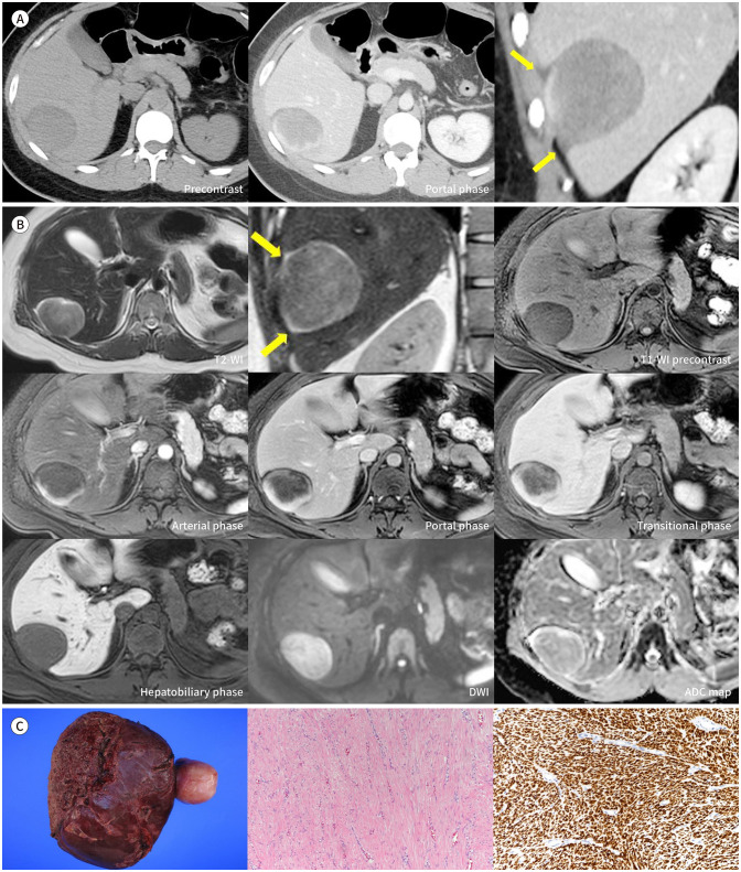
Leiomyoma is a common benign tumor from smooth muscle cells, mostly in the uterus. Peritoneal leiomyomas (PLs) are extremely rare and mostly reported as disseminated peritoneal leiomyomatosis. However, to the best of out knowledge, radiologic findings of isolated PL are not reported in English literature. Herein, we introduce the radiologic findings of PL mimicking hepatic mass in a 34-year-old female. CT showed a mass with curvilinear heterogeneous enhancement at the liver's peripheral area. On MRI, the mass showed gradual and heterogeneous enhancement on gadoxetic acid-enhanced MRI and diffusion restriction. The radiologic diagnosis was a benign hepatic tumor, such as degenerated hemangioma, adenoma, and inflammatory myofibroblastic tumor; however, the mass was diagnosed as PL pathologically.


 分享
分享
 求助内容:
求助内容: 应助结果提醒方式:
 扫码关注我们
