M2 macrophage-derived extracellular vesicles augment immune evasion and development of colorectal cancer via a circRNA_CCDC66/microRNA-342-3p/metadherin axis.
{"title":"M2 macrophage-derived extracellular vesicles augment immune evasion and development of colorectal cancer via a circRNA_CCDC66/microRNA-342-3p/metadherin axis.","authors":"Linfeng Fan, Guofeng Xu, Xiangfu Zeng","doi":"10.1007/s10616-023-00577-z","DOIUrl":null,"url":null,"abstract":"<p><p>The M2 macrophages are major components in the tumor microenvironment and are closely linked to immune suppression and tumor metastasis. This work focuses on how M2 macrophage-derived extracellular vesicles (EVs) affect colorectal cancer (CRC) progression. THP-1 monocytes were induced to differentiate to M0 or M2 macrophages, and the macrophage-derived EVs (M0-EVs and M2-EVs, respectively) were collected and identified. The M2-EVs stimulation augmented proliferation, mobility, and the in vivo tumorigenic activity of CRC cells. Circular RNA_CCDC66 (circ_CCDC66) was highly enriched in M2-EVs and could be delivered into CRC cells. The RNA pull-down and luciferase assays showed that circ_CCDC66 could competitively bind to microRNA (miR)-342-3p, therefore restoring the expression of metadherin (MTDH) mRNA, a target transcript of miR-342-3p. Suppression of circ_CCDC66 in the M2-EVs or specific knockdown of MTDH in CRC significantly blocked the growth and mobility of CRC cells. However, miR-342-3p inhibition restored the malignant phenotype of cancer cells. Moreover, the MTDH knockdown was found to increase the cytotoxicity of CD8<sup>+</sup> T and reduce the protein level of the immune checkpoint PDL1 in CRC cells. In summary, this study reveals that the M2-EVs augment immune evasion and development of CRC by delivering circ_CCDC66 and restoring the MTDH level.</p>","PeriodicalId":10890,"journal":{"name":"Cytotechnology","volume":"75 4","pages":"293-308"},"PeriodicalIF":1.7000,"publicationDate":"2023-08-01","publicationTypes":"Journal Article","fieldsOfStudy":null,"isOpenAccess":false,"openAccessPdf":"https://www.ncbi.nlm.nih.gov/pmc/articles/PMC10299985/pdf/","citationCount":"0","resultStr":null,"platform":"Semanticscholar","paperid":null,"PeriodicalName":"Cytotechnology","FirstCategoryId":"99","ListUrlMain":"https://doi.org/10.1007/s10616-023-00577-z","RegionNum":4,"RegionCategory":"生物学","ArticlePicture":[],"TitleCN":null,"AbstractTextCN":null,"PMCID":null,"EPubDate":"2023/4/19 0:00:00","PubModel":"Epub","JCR":"Q3","JCRName":"BIOTECHNOLOGY & APPLIED MICROBIOLOGY","Score":null,"Total":0}
引用次数: 0
Abstract
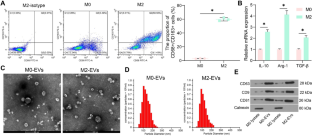
The M2 macrophages are major components in the tumor microenvironment and are closely linked to immune suppression and tumor metastasis. This work focuses on how M2 macrophage-derived extracellular vesicles (EVs) affect colorectal cancer (CRC) progression. THP-1 monocytes were induced to differentiate to M0 or M2 macrophages, and the macrophage-derived EVs (M0-EVs and M2-EVs, respectively) were collected and identified. The M2-EVs stimulation augmented proliferation, mobility, and the in vivo tumorigenic activity of CRC cells. Circular RNA_CCDC66 (circ_CCDC66) was highly enriched in M2-EVs and could be delivered into CRC cells. The RNA pull-down and luciferase assays showed that circ_CCDC66 could competitively bind to microRNA (miR)-342-3p, therefore restoring the expression of metadherin (MTDH) mRNA, a target transcript of miR-342-3p. Suppression of circ_CCDC66 in the M2-EVs or specific knockdown of MTDH in CRC significantly blocked the growth and mobility of CRC cells. However, miR-342-3p inhibition restored the malignant phenotype of cancer cells. Moreover, the MTDH knockdown was found to increase the cytotoxicity of CD8+ T and reduce the protein level of the immune checkpoint PDL1 in CRC cells. In summary, this study reveals that the M2-EVs augment immune evasion and development of CRC by delivering circ_CCDC66 and restoring the MTDH level.
期刊介绍:
The scope of the Journal includes:
1. The derivation, genetic modification and characterization of cell lines, genetic and phenotypic regulation, control of cellular metabolism, cell physiology and biochemistry related to cell function, performance and expression of cell products.
2. Cell culture techniques, substrates, environmental requirements and optimization, cloning, hybridization and molecular biology, including genomic and proteomic tools.
3. Cell culture systems, processes, reactors, scale-up, and industrial production. Descriptions of the design or construction of equipment, media or quality control procedures, that are ancillary to cellular research.
4. The application of animal/human cells in research in the field of stem cell research including maintenance of stemness, differentiation, genetics, and senescence, cancer research, research in immunology, as well as applications in tissue engineering and gene therapy.
5. The use of cell cultures as a substrate for bioassays, biomedical applications and in particular as a replacement for animal models.


 分享
分享
 求助内容:
求助内容: 应助结果提醒方式:
 扫码关注我们
