{"title":"Classification of Aβ State From Brain Amyloid PET Images Using Machine Learning Algorithm.","authors":"Chanda Simfukwe, Reeree Lee, Young Chul Youn","doi":"10.12779/dnd.2023.22.2.61","DOIUrl":null,"url":null,"abstract":"<p><strong>Background and purpose: </strong>Analyzing brain amyloid positron emission tomography (PET) images to access the occurrence of β-amyloid (Aβ) deposition in Alzheimer's patients requires much time and effort from physicians, while the variation of each interpreter may differ. For these reasons, a machine learning model was developed using a convolutional neural network (CNN) as an objective decision to classify the Aβ positive and Aβ negative status from brain amyloid PET images.</p><p><strong>Methods: </strong>A total of 7,344 PET images of 144 subjects were used in this study. The 18F-florbetaben PET was administered to all participants, and the criteria for differentiating Aβ positive and Aβ negative state was based on brain amyloid plaque load score (BAPL) that depended on the visual assessment of PET images by the physicians. We applied the CNN algorithm trained in batches of 51 PET images per subject directory from 2 classes: Aβ positive and Aβ negative states, based on the BAPL scores.</p><p><strong>Results: </strong>The binary classification of the model average performance matrices was evaluated after 40 epochs of three trials based on test datasets. The model accuracy for classifying Aβ positivity and Aβ negativity was (95.00±0.02) in the test dataset. The sensitivity and specificity were (96.00±0.02) and (94.00±0.02), respectively, with an area under the curve of (87.00±0.03).</p><p><strong>Conclusions: </strong>Based on this study, the designed CNN model has the potential to be used clinically to screen amyloid PET images.</p>","PeriodicalId":72779,"journal":{"name":"Dementia and neurocognitive disorders","volume":"22 2","pages":"61-68"},"PeriodicalIF":0.0000,"publicationDate":"2023-04-01","publicationTypes":"Journal Article","fieldsOfStudy":null,"isOpenAccess":false,"openAccessPdf":"https://ftp.ncbi.nlm.nih.gov/pub/pmc/oa_pdf/c9/2f/dnd-22-61.PMC10166673.pdf","citationCount":"0","resultStr":null,"platform":"Semanticscholar","paperid":null,"PeriodicalName":"Dementia and neurocognitive disorders","FirstCategoryId":"1085","ListUrlMain":"https://doi.org/10.12779/dnd.2023.22.2.61","RegionNum":0,"RegionCategory":null,"ArticlePicture":[],"TitleCN":null,"AbstractTextCN":null,"PMCID":null,"EPubDate":"2023/4/30 0:00:00","PubModel":"Epub","JCR":"","JCRName":"","Score":null,"Total":0}
引用次数: 0
Abstract
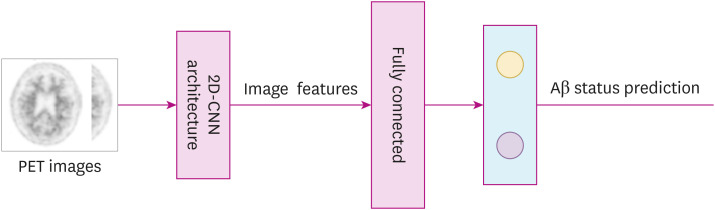
Background and purpose: Analyzing brain amyloid positron emission tomography (PET) images to access the occurrence of β-amyloid (Aβ) deposition in Alzheimer's patients requires much time and effort from physicians, while the variation of each interpreter may differ. For these reasons, a machine learning model was developed using a convolutional neural network (CNN) as an objective decision to classify the Aβ positive and Aβ negative status from brain amyloid PET images.
Methods: A total of 7,344 PET images of 144 subjects were used in this study. The 18F-florbetaben PET was administered to all participants, and the criteria for differentiating Aβ positive and Aβ negative state was based on brain amyloid plaque load score (BAPL) that depended on the visual assessment of PET images by the physicians. We applied the CNN algorithm trained in batches of 51 PET images per subject directory from 2 classes: Aβ positive and Aβ negative states, based on the BAPL scores.
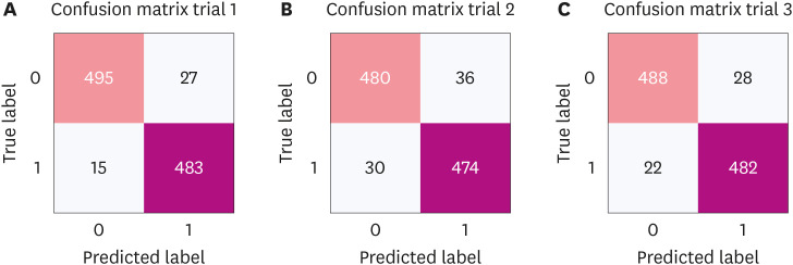
Results: The binary classification of the model average performance matrices was evaluated after 40 epochs of three trials based on test datasets. The model accuracy for classifying Aβ positivity and Aβ negativity was (95.00±0.02) in the test dataset. The sensitivity and specificity were (96.00±0.02) and (94.00±0.02), respectively, with an area under the curve of (87.00±0.03).
Conclusions: Based on this study, the designed CNN model has the potential to be used clinically to screen amyloid PET images.


 分享
分享

 求助内容:
求助内容: 应助结果提醒方式:
 扫码关注我们
