{"title":"The morphogenesis of the rabbit meibomian gland in relation to sex hormones: Immunohistochemical and transmission electron microscopy studies.","authors":"Sara M M El-Desoky, Nada Abdellah","doi":"10.1186/s40850-022-00149-2","DOIUrl":null,"url":null,"abstract":"<p><p>Rabbits have been proposed as a model for the human meibomian gland (MG), a large specific sebaceous gland in the eyelid that consists of secretory acini arranged laterally and related to the central duct via short ductules, with the central duct continuing as an excretory duct to open at the free margin of the lid. First detected at embryonic day 18 as an aggregation of mesenchymal cells in the tarsal plate, it completes its development approximately 2 weeks postnatal when the separation of the eyelids is completed. The Transmission electron microscopy supports the meibocytes' gradient maturation to the meibum's synthesis. While the differentiating cells, their cytoplasm, are well packed with lipid droplets, the basal cells are characterized by a high nuclear to cytoplasm ratio. The androgen and estrogen receptor proteins are expressed in the basal cell and the meibocytes, and increase in age increases in the expression of these proteins. Additionally, the cytokeratin (CK14) is expressed in the basal and differentiating cells of the acini and the ductal epithelium. Therefore, the duct cells of the MG are common in all stem cells. These data concluded that the MG plays a major role in maintaining the health of the ocular surface and preservation of visual acuity. Any abnormalities in the structure of the MG lead to its dysfunction and changes in lipid secretion.</p>","PeriodicalId":48590,"journal":{"name":"BMC Zoology","volume":"7 1","pages":"46"},"PeriodicalIF":1.7000,"publicationDate":"2022-08-11","publicationTypes":"Journal Article","fieldsOfStudy":null,"isOpenAccess":false,"openAccessPdf":"https://www.ncbi.nlm.nih.gov/pmc/articles/PMC10127434/pdf/","citationCount":"1","resultStr":null,"platform":"Semanticscholar","paperid":null,"PeriodicalName":"BMC Zoology","FirstCategoryId":"99","ListUrlMain":"https://doi.org/10.1186/s40850-022-00149-2","RegionNum":3,"RegionCategory":"生物学","ArticlePicture":[],"TitleCN":null,"AbstractTextCN":null,"PMCID":null,"EPubDate":"","PubModel":"","JCR":"Q2","JCRName":"ZOOLOGY","Score":null,"Total":0}
引用次数: 1
Abstract
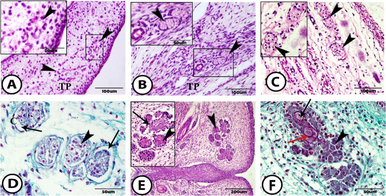
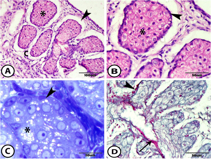
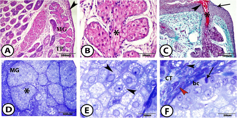
Rabbits have been proposed as a model for the human meibomian gland (MG), a large specific sebaceous gland in the eyelid that consists of secretory acini arranged laterally and related to the central duct via short ductules, with the central duct continuing as an excretory duct to open at the free margin of the lid. First detected at embryonic day 18 as an aggregation of mesenchymal cells in the tarsal plate, it completes its development approximately 2 weeks postnatal when the separation of the eyelids is completed. The Transmission electron microscopy supports the meibocytes' gradient maturation to the meibum's synthesis. While the differentiating cells, their cytoplasm, are well packed with lipid droplets, the basal cells are characterized by a high nuclear to cytoplasm ratio. The androgen and estrogen receptor proteins are expressed in the basal cell and the meibocytes, and increase in age increases in the expression of these proteins. Additionally, the cytokeratin (CK14) is expressed in the basal and differentiating cells of the acini and the ductal epithelium. Therefore, the duct cells of the MG are common in all stem cells. These data concluded that the MG plays a major role in maintaining the health of the ocular surface and preservation of visual acuity. Any abnormalities in the structure of the MG lead to its dysfunction and changes in lipid secretion.
BMC ZoologyAgricultural and Biological Sciences-Animal Science and Zoology
CiteScore
2.30
自引率
6.20%
发文量
53
审稿时长
24 weeks
期刊介绍:
BMC Zoology is an open access, peer-reviewed journal that considers articles on all aspects of zoology, including physiology, mechanistic and functional studies, anatomy, life history, behavior, signalling and communication, cognition, parasitism, taxonomy and conservation.


 分享
分享
 求助内容:
求助内容: 应助结果提醒方式:
 扫码关注我们
