Amir Vokshoor, Harseerat Jajj, Tiffany Grunwald, Steven Kolker, Jack Petros
{"title":"Posterior cervical congenital dermal sinus tract: case report and review of literature.","authors":"Amir Vokshoor, Harseerat Jajj, Tiffany Grunwald, Steven Kolker, Jack Petros","doi":"10.1038/s41394-023-00575-5","DOIUrl":null,"url":null,"abstract":"<p><strong>Background and importance: </strong>Congenital dermal sinus tract (DST) is a rare spinal dysraphism characterized by a persistent tract lined by epithelial cells, beginning at the epidermis and terminating in deeper tissue layers. With 1% of all congenital DST cases found in the cervical region, only 4% of all cases are diagnosed after the age of 20.</p><p><strong>Clinical presentation: </strong>In this case, a 65-year-old woman with a congenital DST at the cervical level presented with symptoms of neck and some arm pain, suboccipital headaches, and unique external characteristics. Neck Disability Index and visual analog scale were used to assess the patient's preoperative and postoperative pain, and quality of life. Patient underwent an operative intervention, where the DST was surgically removed followed by interlaminar decompression at C1-C2, excision of the epidural component, and biopsy followed by plastic surgical repair. Pathology analysis indicated a squamous epithelial-lined sinus tract interacting with the dura. Most notably, a meningothelial proliferation with associated psammomatous calcifications was identified, similar to a meningioma.</p><p><strong>Conclusion: </strong>A review of literature was conducted to further discuss clinical and radiological presentation as well as to document the novel appearance of this congenital DST. As one of the oldest cases of DST, it demonstrated unusual pathological characteristics with a meningothelial proliferation, compatible with meningioma, reported at the epidural level.</p>","PeriodicalId":22079,"journal":{"name":"Spinal Cord Series and Cases","volume":"9 1","pages":"40"},"PeriodicalIF":0.9000,"publicationDate":"2023-08-02","publicationTypes":"Journal Article","fieldsOfStudy":null,"isOpenAccess":false,"openAccessPdf":"https://www.ncbi.nlm.nih.gov/pmc/articles/PMC10397304/pdf/","citationCount":"0","resultStr":null,"platform":"Semanticscholar","paperid":null,"PeriodicalName":"Spinal Cord Series and Cases","FirstCategoryId":"1085","ListUrlMain":"https://doi.org/10.1038/s41394-023-00575-5","RegionNum":0,"RegionCategory":null,"ArticlePicture":[],"TitleCN":null,"AbstractTextCN":null,"PMCID":null,"EPubDate":"","PubModel":"","JCR":"Q4","JCRName":"CLINICAL NEUROLOGY","Score":null,"Total":0}
引用次数: 0
Abstract
Background and importance: Congenital dermal sinus tract (DST) is a rare spinal dysraphism characterized by a persistent tract lined by epithelial cells, beginning at the epidermis and terminating in deeper tissue layers. With 1% of all congenital DST cases found in the cervical region, only 4% of all cases are diagnosed after the age of 20.
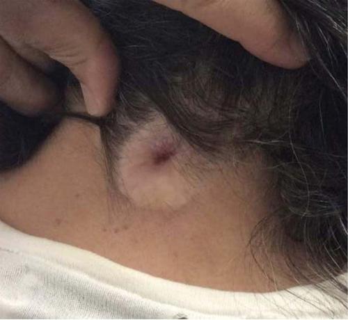
Clinical presentation: In this case, a 65-year-old woman with a congenital DST at the cervical level presented with symptoms of neck and some arm pain, suboccipital headaches, and unique external characteristics. Neck Disability Index and visual analog scale were used to assess the patient's preoperative and postoperative pain, and quality of life. Patient underwent an operative intervention, where the DST was surgically removed followed by interlaminar decompression at C1-C2, excision of the epidural component, and biopsy followed by plastic surgical repair. Pathology analysis indicated a squamous epithelial-lined sinus tract interacting with the dura. Most notably, a meningothelial proliferation with associated psammomatous calcifications was identified, similar to a meningioma.
Conclusion: A review of literature was conducted to further discuss clinical and radiological presentation as well as to document the novel appearance of this congenital DST. As one of the oldest cases of DST, it demonstrated unusual pathological characteristics with a meningothelial proliferation, compatible with meningioma, reported at the epidural level.


 分享
分享
 求助内容:
求助内容: 应助结果提醒方式:
 扫码关注我们
