{"title":"A rare find of a maxillary third molar with five roots: a case report of an unusual morphology.","authors":"Oana Cella Andrei, Gabriela Ciavoi, Magdalena Natalia Dina, Liana Todor, Daniela Ioana Tărlungeanu, Ruxandra Mărgărit","doi":"10.47162/RJME.64.2.19","DOIUrl":null,"url":null,"abstract":"<p><p>Third molars are frequently absent or impacted; their unpredictable prevalence and morphology, with frequent anatomical variations, is a challenge for the practitioner. The number of roots of the upper third molars vary from one to five, but five roots upper third molars' cases are extremely rare. The case reported here is of a normotrophic male patient, with no significant general medical history, having extracted an impacted maxillary upper molar, with an unusual position and dimensions and five completely separated formed roots, orientated in diverse directions. It highlights the importance of using the correct surgical technique, without excessive pressure, for avoiding the fracture of the maxillary tuberosity, the displacement of the molar into the pterygopalatine fossa or the opening of the maxillary sinus, especially in the absence of good quality imagistic information like cone-beam computed tomography. The case presented in this paper is very rare and highlights the significance of anatomical knowledge, which is mandatory for managing these cases, to avoid trauma, accidents, and complications and to maintain an optimal time of extraction, since such variations occur, and the conventional radiographs do not offer enough data to clarify the diagnosis.</p>","PeriodicalId":54447,"journal":{"name":"Romanian Journal of Morphology and Embryology","volume":"64 2","pages":"275-278"},"PeriodicalIF":1.5000,"publicationDate":"2023-04-01","publicationTypes":"Journal Article","fieldsOfStudy":null,"isOpenAccess":false,"openAccessPdf":"https://ftp.ncbi.nlm.nih.gov/pub/pmc/oa_pdf/2e/24/RJME-64-2-275.PMC10520373.pdf","citationCount":"0","resultStr":null,"platform":"Semanticscholar","paperid":null,"PeriodicalName":"Romanian Journal of Morphology and Embryology","FirstCategoryId":"3","ListUrlMain":"https://doi.org/10.47162/RJME.64.2.19","RegionNum":4,"RegionCategory":"医学","ArticlePicture":[],"TitleCN":null,"AbstractTextCN":null,"PMCID":null,"EPubDate":"","PubModel":"","JCR":"Q4","JCRName":"DEVELOPMENTAL BIOLOGY","Score":null,"Total":0}
引用次数: 0
Abstract
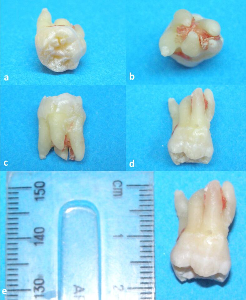
Third molars are frequently absent or impacted; their unpredictable prevalence and morphology, with frequent anatomical variations, is a challenge for the practitioner. The number of roots of the upper third molars vary from one to five, but five roots upper third molars' cases are extremely rare. The case reported here is of a normotrophic male patient, with no significant general medical history, having extracted an impacted maxillary upper molar, with an unusual position and dimensions and five completely separated formed roots, orientated in diverse directions. It highlights the importance of using the correct surgical technique, without excessive pressure, for avoiding the fracture of the maxillary tuberosity, the displacement of the molar into the pterygopalatine fossa or the opening of the maxillary sinus, especially in the absence of good quality imagistic information like cone-beam computed tomography. The case presented in this paper is very rare and highlights the significance of anatomical knowledge, which is mandatory for managing these cases, to avoid trauma, accidents, and complications and to maintain an optimal time of extraction, since such variations occur, and the conventional radiographs do not offer enough data to clarify the diagnosis.
期刊介绍:
Romanian Journal of Morphology and Embryology (Rom J Morphol Embryol) publishes studies on all aspects of normal morphology and human comparative and experimental pathology. The Journal accepts only researches that utilize modern investigation methods (studies of anatomy, pathology, cytopathology, immunohistochemistry, histochemistry, immunology, morphometry, molecular and cellular biology, electronic microscopy, etc.).


 分享
分享
 求助内容:
求助内容: 应助结果提醒方式:
 扫码关注我们
