María A Moyaho-Bernal, Rosendo Carrasco-Gutiérrez, Rosario Jiménez-Flores, Gladis Juárez-Luna, Gerardo R López-Del Pino, Luz V Mendoza-García, Bernardo Teutle-Coyotecatl
{"title":"三根原发性下颌第一和第二磨牙的患病率:墨西哥人群的临床和放射学发现。","authors":"María A Moyaho-Bernal, Rosendo Carrasco-Gutiérrez, Rosario Jiménez-Flores, Gladis Juárez-Luna, Gerardo R López-Del Pino, Luz V Mendoza-García, Bernardo Teutle-Coyotecatl","doi":"10.54589/aol.34/2/149","DOIUrl":null,"url":null,"abstract":"<p><p>The aim of this study was to determine prevalence and describe the clinical and radiographic findings of three-rooted primary mandibular first and second molars in a Mexican population. Intraoral periapical radiograph, orthopantomogram or cone beam computed tomography (CBCT) were obtained. A total 2284 children from the state of Puebla, Mexico were examined, of whom 20 presented an anatomic variant in tooth crown shape. Of the total teeth with crown alterations, 10 first and 5 second primary mandibular molars were found to have supernumerary roots. In one case, it was possible to obtain micro-CT images. The study recorded prevalence, unilateral or bilateral occurrence, and ratio between sexes. Data were analyzed using descriptive statistics. Clinical findings were presence of an anatomical variation (tuberculum paramolare / right and/or left cervical convexity) in primary mandibular first molars. Second molars presented conventional crown morphology. Prevalence of three-rooted primary mandibular first and second molars was 0.44% and 0.22%, respectively. Male: female ratio for presence of threerooted primary mandibular first molars was 4:1, showing genetic predisposition in males, and for second molars it was 1.5:1, with no predisposition according to sex. The clinical and radiographic anatomical variants in primary molars should be considered by pediatric dentists during routine care because they may cause difficulties in restorations.</p>","PeriodicalId":7033,"journal":{"name":"Acta odontologica latinoamericana : AOL","volume":"34 2","pages":"149-155"},"PeriodicalIF":0.0000,"publicationDate":"2021-08-01","publicationTypes":"Journal Article","fieldsOfStudy":null,"isOpenAccess":false,"openAccessPdf":"https://ftp.ncbi.nlm.nih.gov/pub/pmc/oa_pdf/be/45/1852-4834-34-2-149.PMC10315086.pdf","citationCount":"2","resultStr":"{\"title\":\"Prevalence of three-rooted primary mandibular first and second molars: clinical and radiographic findings in a Mexican population.\",\"authors\":\"María A Moyaho-Bernal, Rosendo Carrasco-Gutiérrez, Rosario Jiménez-Flores, Gladis Juárez-Luna, Gerardo R López-Del Pino, Luz V Mendoza-García, Bernardo Teutle-Coyotecatl\",\"doi\":\"10.54589/aol.34/2/149\",\"DOIUrl\":null,\"url\":null,\"abstract\":\"<p><p>The aim of this study was to determine prevalence and describe the clinical and radiographic findings of three-rooted primary mandibular first and second molars in a Mexican population. Intraoral periapical radiograph, orthopantomogram or cone beam computed tomography (CBCT) were obtained. A total 2284 children from the state of Puebla, Mexico were examined, of whom 20 presented an anatomic variant in tooth crown shape. Of the total teeth with crown alterations, 10 first and 5 second primary mandibular molars were found to have supernumerary roots. In one case, it was possible to obtain micro-CT images. The study recorded prevalence, unilateral or bilateral occurrence, and ratio between sexes. Data were analyzed using descriptive statistics. Clinical findings were presence of an anatomical variation (tuberculum paramolare / right and/or left cervical convexity) in primary mandibular first molars. Second molars presented conventional crown morphology. Prevalence of three-rooted primary mandibular first and second molars was 0.44% and 0.22%, respectively. Male: female ratio for presence of threerooted primary mandibular first molars was 4:1, showing genetic predisposition in males, and for second molars it was 1.5:1, with no predisposition according to sex. The clinical and radiographic anatomical variants in primary molars should be considered by pediatric dentists during routine care because they may cause difficulties in restorations.</p>\",\"PeriodicalId\":7033,\"journal\":{\"name\":\"Acta odontologica latinoamericana : AOL\",\"volume\":\"34 2\",\"pages\":\"149-155\"},\"PeriodicalIF\":0.0000,\"publicationDate\":\"2021-08-01\",\"publicationTypes\":\"Journal Article\",\"fieldsOfStudy\":null,\"isOpenAccess\":false,\"openAccessPdf\":\"https://ftp.ncbi.nlm.nih.gov/pub/pmc/oa_pdf/be/45/1852-4834-34-2-149.PMC10315086.pdf\",\"citationCount\":\"2\",\"resultStr\":null,\"platform\":\"Semanticscholar\",\"paperid\":null,\"PeriodicalName\":\"Acta odontologica latinoamericana : AOL\",\"FirstCategoryId\":\"1085\",\"ListUrlMain\":\"https://doi.org/10.54589/aol.34/2/149\",\"RegionNum\":0,\"RegionCategory\":null,\"ArticlePicture\":[],\"TitleCN\":null,\"AbstractTextCN\":null,\"PMCID\":null,\"EPubDate\":\"\",\"PubModel\":\"\",\"JCR\":\"\",\"JCRName\":\"\",\"Score\":null,\"Total\":0}","platform":"Semanticscholar","paperid":null,"PeriodicalName":"Acta odontologica latinoamericana : AOL","FirstCategoryId":"1085","ListUrlMain":"https://doi.org/10.54589/aol.34/2/149","RegionNum":0,"RegionCategory":null,"ArticlePicture":[],"TitleCN":null,"AbstractTextCN":null,"PMCID":null,"EPubDate":"","PubModel":"","JCR":"","JCRName":"","Score":null,"Total":0}
Prevalence of three-rooted primary mandibular first and second molars: clinical and radiographic findings in a Mexican population.
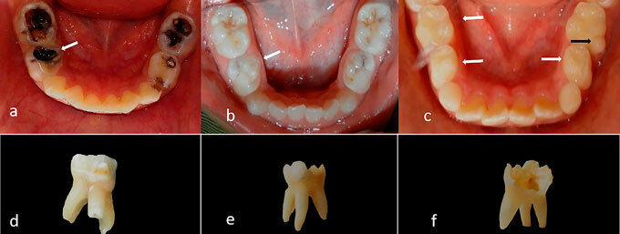
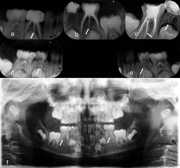
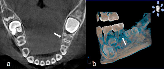
The aim of this study was to determine prevalence and describe the clinical and radiographic findings of three-rooted primary mandibular first and second molars in a Mexican population. Intraoral periapical radiograph, orthopantomogram or cone beam computed tomography (CBCT) were obtained. A total 2284 children from the state of Puebla, Mexico were examined, of whom 20 presented an anatomic variant in tooth crown shape. Of the total teeth with crown alterations, 10 first and 5 second primary mandibular molars were found to have supernumerary roots. In one case, it was possible to obtain micro-CT images. The study recorded prevalence, unilateral or bilateral occurrence, and ratio between sexes. Data were analyzed using descriptive statistics. Clinical findings were presence of an anatomical variation (tuberculum paramolare / right and/or left cervical convexity) in primary mandibular first molars. Second molars presented conventional crown morphology. Prevalence of three-rooted primary mandibular first and second molars was 0.44% and 0.22%, respectively. Male: female ratio for presence of threerooted primary mandibular first molars was 4:1, showing genetic predisposition in males, and for second molars it was 1.5:1, with no predisposition according to sex. The clinical and radiographic anatomical variants in primary molars should be considered by pediatric dentists during routine care because they may cause difficulties in restorations.


 分享
分享

 求助内容:
求助内容: 应助结果提醒方式:
 扫码关注我们
