Andrea Taddeucci, Francesco Zinna, Giuliano Siligardi and Lorenzo Di Bari*,
{"title":"手性有机染料薄膜的圆偏振显微镜研究","authors":"Andrea Taddeucci, Francesco Zinna, Giuliano Siligardi and Lorenzo Di Bari*, ","doi":"10.1021/cbmi.3c00049","DOIUrl":null,"url":null,"abstract":"<p >We introduce an optical microscopy technique, circularly polarized microscopy or CPM, able to afford spatially resolved electronic circular dichroism (ECD) of thin films of chiral organic semiconductors through a commercial microscope equipped with a camera and inexpensive optics. Provided the dichroic ratio is sufficiently large, the spatial resolution is on the order of the μm and is only limited by the magnification optics integrated in the microscope. We apply CPM to thin films of small chiral π-conjugated molecules, which gave rise to ordered aggregates in the thin layer. Primarily, conventional ECD can reveal and characterize chiral supramolecular structures and possible interferences between anisotropic properties of solid samples; however, it cannot generally account for the spatial distribution of such properties. CPM offers a characterization of supramolecular chirality and of commingling polarization anisotropies of the material, describing their local distribution. To validate CPM, we demonstrated that it can be adopted to quantify the local ECD of samples characterized by intense signals, virtually on any standard optical microscope.</p>","PeriodicalId":53181,"journal":{"name":"Chemical & Biomedical Imaging","volume":"1 5","pages":"471–478"},"PeriodicalIF":5.7000,"publicationDate":"2023-06-09","publicationTypes":"Journal Article","fieldsOfStudy":null,"isOpenAccess":false,"openAccessPdf":"https://ftp.ncbi.nlm.nih.gov/pub/pmc/oa_pdf/3b/fc/im3c00049.PMC10467535.pdf","citationCount":"0","resultStr":"{\"title\":\"Circularly Polarized Microscopy of Thin Films of Chiral Organic Dyes\",\"authors\":\"Andrea Taddeucci, Francesco Zinna, Giuliano Siligardi and Lorenzo Di Bari*, \",\"doi\":\"10.1021/cbmi.3c00049\",\"DOIUrl\":null,\"url\":null,\"abstract\":\"<p >We introduce an optical microscopy technique, circularly polarized microscopy or CPM, able to afford spatially resolved electronic circular dichroism (ECD) of thin films of chiral organic semiconductors through a commercial microscope equipped with a camera and inexpensive optics. Provided the dichroic ratio is sufficiently large, the spatial resolution is on the order of the μm and is only limited by the magnification optics integrated in the microscope. We apply CPM to thin films of small chiral π-conjugated molecules, which gave rise to ordered aggregates in the thin layer. Primarily, conventional ECD can reveal and characterize chiral supramolecular structures and possible interferences between anisotropic properties of solid samples; however, it cannot generally account for the spatial distribution of such properties. CPM offers a characterization of supramolecular chirality and of commingling polarization anisotropies of the material, describing their local distribution. To validate CPM, we demonstrated that it can be adopted to quantify the local ECD of samples characterized by intense signals, virtually on any standard optical microscope.</p>\",\"PeriodicalId\":53181,\"journal\":{\"name\":\"Chemical & Biomedical Imaging\",\"volume\":\"1 5\",\"pages\":\"471–478\"},\"PeriodicalIF\":5.7000,\"publicationDate\":\"2023-06-09\",\"publicationTypes\":\"Journal Article\",\"fieldsOfStudy\":null,\"isOpenAccess\":false,\"openAccessPdf\":\"https://ftp.ncbi.nlm.nih.gov/pub/pmc/oa_pdf/3b/fc/im3c00049.PMC10467535.pdf\",\"citationCount\":\"0\",\"resultStr\":null,\"platform\":\"Semanticscholar\",\"paperid\":null,\"PeriodicalName\":\"Chemical & Biomedical Imaging\",\"FirstCategoryId\":\"1085\",\"ListUrlMain\":\"https://pubs.acs.org/doi/10.1021/cbmi.3c00049\",\"RegionNum\":0,\"RegionCategory\":null,\"ArticlePicture\":[],\"TitleCN\":null,\"AbstractTextCN\":null,\"PMCID\":null,\"EPubDate\":\"\",\"PubModel\":\"\",\"JCR\":\"\",\"JCRName\":\"\",\"Score\":null,\"Total\":0}","platform":"Semanticscholar","paperid":null,"PeriodicalName":"Chemical & Biomedical Imaging","FirstCategoryId":"1085","ListUrlMain":"https://pubs.acs.org/doi/10.1021/cbmi.3c00049","RegionNum":0,"RegionCategory":null,"ArticlePicture":[],"TitleCN":null,"AbstractTextCN":null,"PMCID":null,"EPubDate":"","PubModel":"","JCR":"","JCRName":"","Score":null,"Total":0}
Circularly Polarized Microscopy of Thin Films of Chiral Organic Dyes
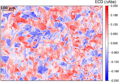
We introduce an optical microscopy technique, circularly polarized microscopy or CPM, able to afford spatially resolved electronic circular dichroism (ECD) of thin films of chiral organic semiconductors through a commercial microscope equipped with a camera and inexpensive optics. Provided the dichroic ratio is sufficiently large, the spatial resolution is on the order of the μm and is only limited by the magnification optics integrated in the microscope. We apply CPM to thin films of small chiral π-conjugated molecules, which gave rise to ordered aggregates in the thin layer. Primarily, conventional ECD can reveal and characterize chiral supramolecular structures and possible interferences between anisotropic properties of solid samples; however, it cannot generally account for the spatial distribution of such properties. CPM offers a characterization of supramolecular chirality and of commingling polarization anisotropies of the material, describing their local distribution. To validate CPM, we demonstrated that it can be adopted to quantify the local ECD of samples characterized by intense signals, virtually on any standard optical microscope.
期刊介绍:
Chemical & Biomedical Imaging is a peer-reviewed open access journal devoted to the publication of cutting-edge research papers on all aspects of chemical and biomedical imaging. This interdisciplinary field sits at the intersection of chemistry physics biology materials engineering and medicine. The journal aims to bring together researchers from across these disciplines to address cutting-edge challenges of fundamental research and applications.Topics of particular interest include but are not limited to:Imaging of processes and reactionsImaging of nanoscale microscale and mesoscale materialsImaging of biological interactions and interfacesSingle-molecule and cellular imagingWhole-organ and whole-body imagingMolecular imaging probes and contrast agentsBioluminescence chemiluminescence and electrochemiluminescence imagingNanophotonics and imagingChemical tools for new imaging modalitiesChemical and imaging techniques in diagnosis and therapyImaging-guided drug deliveryAI and machine learning assisted imaging


 分享
分享
 求助内容:
求助内容: 应助结果提醒方式:
 扫码关注我们
