Saber Saharkhiz, Zohreh Abdolmaleki, Mohammad Amin Eslampour
{"title":"透明质酸/硅纳米颗粒支架可诱导移植到附睾脂肪组织的小鼠精原干细胞增殖和分化。","authors":"Saber Saharkhiz, Zohreh Abdolmaleki, Mohammad Amin Eslampour","doi":"10.1007/s10561-023-10093-1","DOIUrl":null,"url":null,"abstract":"<p><p>Spermatogonia stem cells (SSCs) are a unique cell population maintaining male spermatogenesis during life, through their potential for proliferation and differentiation. The application of silicon nanoparticles (SNs) and hyaluronic acid (HA) to induce the differentiation of SSCs seems promising. Herein, we investigate the effect of SN and HA scaffolds on the progression of SSCs spermatogenesis in mice. Initially SSCs were isolated from healthy immature mice and cultured on prepared scaffolds (HA, SN, and HA/SN) in a 3D culture system. Then viability of SSCs cultured on scaffolds was examined using MTT assay and Acridine Orange staining. Then SSCs cultured on scaffolds were transplanted into epididymal adipose tissue (EAT) in mature mice and the result was studied by H&E and IHC staining 8 weeks after transplantation. MTT and Acridine Orange analysis revealed that among three different scaffolds HA/SN based scaffold causes considerable toxicity on SSCs (P < 0.05) while H&E staining showed that culture of SSCs on HA, SN, and HA/SN scaffolds has a positive effect on the progression of SSCs spermatogenesis after transplantation into EAT. IHC staining identified TP1, TEKT1, and PLZF as crucial biomarkers in the spermatogenesis development of SSCs transplanted to EAT. According to the presence of these biomarkers in different experimental groups, we found the most spermatogenesis development in SSCs cultured on HA/SN scaffold (PLZF, P < 0.01) (TEKT1, P < 0.01) (TP1, P < 0.001). Our study showed that, although the cytotoxic effect of the HA/SN scaffold decreases the viability rate of SSCs; however, SSCs that survive on HA/SN scaffold showed more ability to progress in spermatogenesis after transplantation into EAT.</p>","PeriodicalId":9723,"journal":{"name":"Cell and Tissue Banking","volume":" ","pages":"231-243"},"PeriodicalIF":2.0000,"publicationDate":"2024-03-01","publicationTypes":"Journal Article","fieldsOfStudy":null,"isOpenAccess":false,"openAccessPdf":"","citationCount":"0","resultStr":"{\"title\":\"Hyaluronic acid/silicon nanoparticle scaffold induces proliferation and differentiation of mouse spermatogonial stem cells transplanted to epididymal adipose tissue.\",\"authors\":\"Saber Saharkhiz, Zohreh Abdolmaleki, Mohammad Amin Eslampour\",\"doi\":\"10.1007/s10561-023-10093-1\",\"DOIUrl\":null,\"url\":null,\"abstract\":\"<p><p>Spermatogonia stem cells (SSCs) are a unique cell population maintaining male spermatogenesis during life, through their potential for proliferation and differentiation. The application of silicon nanoparticles (SNs) and hyaluronic acid (HA) to induce the differentiation of SSCs seems promising. Herein, we investigate the effect of SN and HA scaffolds on the progression of SSCs spermatogenesis in mice. Initially SSCs were isolated from healthy immature mice and cultured on prepared scaffolds (HA, SN, and HA/SN) in a 3D culture system. Then viability of SSCs cultured on scaffolds was examined using MTT assay and Acridine Orange staining. Then SSCs cultured on scaffolds were transplanted into epididymal adipose tissue (EAT) in mature mice and the result was studied by H&E and IHC staining 8 weeks after transplantation. MTT and Acridine Orange analysis revealed that among three different scaffolds HA/SN based scaffold causes considerable toxicity on SSCs (P < 0.05) while H&E staining showed that culture of SSCs on HA, SN, and HA/SN scaffolds has a positive effect on the progression of SSCs spermatogenesis after transplantation into EAT. IHC staining identified TP1, TEKT1, and PLZF as crucial biomarkers in the spermatogenesis development of SSCs transplanted to EAT. According to the presence of these biomarkers in different experimental groups, we found the most spermatogenesis development in SSCs cultured on HA/SN scaffold (PLZF, P < 0.01) (TEKT1, P < 0.01) (TP1, P < 0.001). Our study showed that, although the cytotoxic effect of the HA/SN scaffold decreases the viability rate of SSCs; however, SSCs that survive on HA/SN scaffold showed more ability to progress in spermatogenesis after transplantation into EAT.</p>\",\"PeriodicalId\":9723,\"journal\":{\"name\":\"Cell and Tissue Banking\",\"volume\":\" \",\"pages\":\"231-243\"},\"PeriodicalIF\":2.0000,\"publicationDate\":\"2024-03-01\",\"publicationTypes\":\"Journal Article\",\"fieldsOfStudy\":null,\"isOpenAccess\":false,\"openAccessPdf\":\"\",\"citationCount\":\"0\",\"resultStr\":null,\"platform\":\"Semanticscholar\",\"paperid\":null,\"PeriodicalName\":\"Cell and Tissue Banking\",\"FirstCategoryId\":\"5\",\"ListUrlMain\":\"https://doi.org/10.1007/s10561-023-10093-1\",\"RegionNum\":4,\"RegionCategory\":\"医学\",\"ArticlePicture\":[],\"TitleCN\":null,\"AbstractTextCN\":null,\"PMCID\":null,\"EPubDate\":\"2023/9/7 0:00:00\",\"PubModel\":\"Epub\",\"JCR\":\"Q4\",\"JCRName\":\"CELL BIOLOGY\",\"Score\":null,\"Total\":0}","platform":"Semanticscholar","paperid":null,"PeriodicalName":"Cell and Tissue Banking","FirstCategoryId":"5","ListUrlMain":"https://doi.org/10.1007/s10561-023-10093-1","RegionNum":4,"RegionCategory":"医学","ArticlePicture":[],"TitleCN":null,"AbstractTextCN":null,"PMCID":null,"EPubDate":"2023/9/7 0:00:00","PubModel":"Epub","JCR":"Q4","JCRName":"CELL BIOLOGY","Score":null,"Total":0}
Hyaluronic acid/silicon nanoparticle scaffold induces proliferation and differentiation of mouse spermatogonial stem cells transplanted to epididymal adipose tissue.
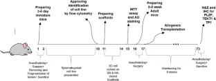
Spermatogonia stem cells (SSCs) are a unique cell population maintaining male spermatogenesis during life, through their potential for proliferation and differentiation. The application of silicon nanoparticles (SNs) and hyaluronic acid (HA) to induce the differentiation of SSCs seems promising. Herein, we investigate the effect of SN and HA scaffolds on the progression of SSCs spermatogenesis in mice. Initially SSCs were isolated from healthy immature mice and cultured on prepared scaffolds (HA, SN, and HA/SN) in a 3D culture system. Then viability of SSCs cultured on scaffolds was examined using MTT assay and Acridine Orange staining. Then SSCs cultured on scaffolds were transplanted into epididymal adipose tissue (EAT) in mature mice and the result was studied by H&E and IHC staining 8 weeks after transplantation. MTT and Acridine Orange analysis revealed that among three different scaffolds HA/SN based scaffold causes considerable toxicity on SSCs (P < 0.05) while H&E staining showed that culture of SSCs on HA, SN, and HA/SN scaffolds has a positive effect on the progression of SSCs spermatogenesis after transplantation into EAT. IHC staining identified TP1, TEKT1, and PLZF as crucial biomarkers in the spermatogenesis development of SSCs transplanted to EAT. According to the presence of these biomarkers in different experimental groups, we found the most spermatogenesis development in SSCs cultured on HA/SN scaffold (PLZF, P < 0.01) (TEKT1, P < 0.01) (TP1, P < 0.001). Our study showed that, although the cytotoxic effect of the HA/SN scaffold decreases the viability rate of SSCs; however, SSCs that survive on HA/SN scaffold showed more ability to progress in spermatogenesis after transplantation into EAT.
期刊介绍:
Cell and Tissue Banking provides a forum for disseminating information to scientists and clinicians involved in the banking and transplantation of cells and tissues. Cell and Tissue Banking is an international, peer-reviewed journal that publishes original papers in the following areas:
basic research concerning general aspects of tissue banking such as quality assurance and control of banked cells/tissues, effects of preservation and sterilisation methods on cells/tissues, biotechnology, etc.; clinical applications of banked cells/tissues; standards of practice in procurement, processing, storage and distribution of cells/tissues; ethical issues; medico-legal issues.


 分享
分享
 求助内容:
求助内容: 应助结果提醒方式:
 扫码关注我们
