Meng Lu, Weixu Li, Yeqing Wang, Lixian Yuan, Meng Cao
{"title":"犬正畸种植体位置转换技术的初步研究。","authors":"Meng Lu, Weixu Li, Yeqing Wang, Lixian Yuan, Meng Cao","doi":"10.1186/s13005-023-00373-2","DOIUrl":null,"url":null,"abstract":"<p><strong>Background: </strong>To evaluate the quantity and quality of bone in the newly formed edentulous area produced by the orthodontic implant site-switching technique.</p><p><strong>Methods: </strong>The bilateral maxillary first premolars of five beagle dogs were extracted and bone defects were created. The right and left sides of the maxilla were randomly divided into control and experimental sides. On the experimental side, the maxillary second premolar was mesially moved into the position of the missing first premolar. On the control side, the second maxillary premolar was extracted. Six months later, the beagles were euthanized. Microcomputer tomography was used to analyze bone microstructure parameters, alveolar bone height and alveolar bone width of the regenerated bone. Histological analysis was performed by staining tissue sections with toluidine blue.</p><p><strong>Results: </strong>Median BV/TV values in the experimental group (81.78%) were significantly larger than those in the control group (35.67%; p = 0.04). Median Tb.Sp values in the experimental group (0.14 mm) were significantly lower than those in the control group (0.54 mm; p = 0.04). Median Tb.Th values in the experimental group (0.48 mm) were significantly higher than those in the control group (0.21 mm; p = 0.04). Median Tb.Pf values in the experimental group (0.65/mm) were significantly lower than those in the control group (3.15/mm; p = 0.04). There was no significant difference in the trabecular number (Tb.N) between the two groups (p = 0.23). The median alveolar bone height values in the experimental group (-0.81 mm) were significantly higher than those in the control group (-2.11 mm; p = 0.04) at a distance 5 mm from the mesial CEJ of the third premolar. The median alveolar bone height values in the experimental group (0.45 mm) were significantly higher than those in the control group (-1.70 mm; p = 0.04) at a distance 6 mm from the mesial CEJ of the third premolar. There was no significant difference in alveolar bone width when compared between the two groups (p > 0.05).</p><p><strong>Conclusions: </strong>The newly formed edentulous area created by orthodontic treatment had more compact and thicker trabeculae than the extraction socket. Furthermore, the newly formed edentulous area had a greater alveolar bone height available for the placement of implants.</p>","PeriodicalId":12994,"journal":{"name":"Head & Face Medicine","volume":null,"pages":null},"PeriodicalIF":2.4000,"publicationDate":"2023-07-14","publicationTypes":"Journal Article","fieldsOfStudy":null,"isOpenAccess":false,"openAccessPdf":"https://www.ncbi.nlm.nih.gov/pmc/articles/PMC10347830/pdf/","citationCount":"0","resultStr":"{\"title\":\"The orthodontic implant site-switching technique: a preliminary study in dogs.\",\"authors\":\"Meng Lu, Weixu Li, Yeqing Wang, Lixian Yuan, Meng Cao\",\"doi\":\"10.1186/s13005-023-00373-2\",\"DOIUrl\":null,\"url\":null,\"abstract\":\"<p><strong>Background: </strong>To evaluate the quantity and quality of bone in the newly formed edentulous area produced by the orthodontic implant site-switching technique.</p><p><strong>Methods: </strong>The bilateral maxillary first premolars of five beagle dogs were extracted and bone defects were created. The right and left sides of the maxilla were randomly divided into control and experimental sides. On the experimental side, the maxillary second premolar was mesially moved into the position of the missing first premolar. On the control side, the second maxillary premolar was extracted. Six months later, the beagles were euthanized. Microcomputer tomography was used to analyze bone microstructure parameters, alveolar bone height and alveolar bone width of the regenerated bone. Histological analysis was performed by staining tissue sections with toluidine blue.</p><p><strong>Results: </strong>Median BV/TV values in the experimental group (81.78%) were significantly larger than those in the control group (35.67%; p = 0.04). Median Tb.Sp values in the experimental group (0.14 mm) were significantly lower than those in the control group (0.54 mm; p = 0.04). Median Tb.Th values in the experimental group (0.48 mm) were significantly higher than those in the control group (0.21 mm; p = 0.04). Median Tb.Pf values in the experimental group (0.65/mm) were significantly lower than those in the control group (3.15/mm; p = 0.04). There was no significant difference in the trabecular number (Tb.N) between the two groups (p = 0.23). The median alveolar bone height values in the experimental group (-0.81 mm) were significantly higher than those in the control group (-2.11 mm; p = 0.04) at a distance 5 mm from the mesial CEJ of the third premolar. The median alveolar bone height values in the experimental group (0.45 mm) were significantly higher than those in the control group (-1.70 mm; p = 0.04) at a distance 6 mm from the mesial CEJ of the third premolar. There was no significant difference in alveolar bone width when compared between the two groups (p > 0.05).</p><p><strong>Conclusions: </strong>The newly formed edentulous area created by orthodontic treatment had more compact and thicker trabeculae than the extraction socket. Furthermore, the newly formed edentulous area had a greater alveolar bone height available for the placement of implants.</p>\",\"PeriodicalId\":12994,\"journal\":{\"name\":\"Head & Face Medicine\",\"volume\":null,\"pages\":null},\"PeriodicalIF\":2.4000,\"publicationDate\":\"2023-07-14\",\"publicationTypes\":\"Journal Article\",\"fieldsOfStudy\":null,\"isOpenAccess\":false,\"openAccessPdf\":\"https://www.ncbi.nlm.nih.gov/pmc/articles/PMC10347830/pdf/\",\"citationCount\":\"0\",\"resultStr\":null,\"platform\":\"Semanticscholar\",\"paperid\":null,\"PeriodicalName\":\"Head & Face Medicine\",\"FirstCategoryId\":\"3\",\"ListUrlMain\":\"https://doi.org/10.1186/s13005-023-00373-2\",\"RegionNum\":2,\"RegionCategory\":\"医学\",\"ArticlePicture\":[],\"TitleCN\":null,\"AbstractTextCN\":null,\"PMCID\":null,\"EPubDate\":\"\",\"PubModel\":\"\",\"JCR\":\"Q2\",\"JCRName\":\"DENTISTRY, ORAL SURGERY & MEDICINE\",\"Score\":null,\"Total\":0}","platform":"Semanticscholar","paperid":null,"PeriodicalName":"Head & Face Medicine","FirstCategoryId":"3","ListUrlMain":"https://doi.org/10.1186/s13005-023-00373-2","RegionNum":2,"RegionCategory":"医学","ArticlePicture":[],"TitleCN":null,"AbstractTextCN":null,"PMCID":null,"EPubDate":"","PubModel":"","JCR":"Q2","JCRName":"DENTISTRY, ORAL SURGERY & MEDICINE","Score":null,"Total":0}
The orthodontic implant site-switching technique: a preliminary study in dogs.
Background: To evaluate the quantity and quality of bone in the newly formed edentulous area produced by the orthodontic implant site-switching technique.
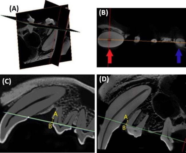
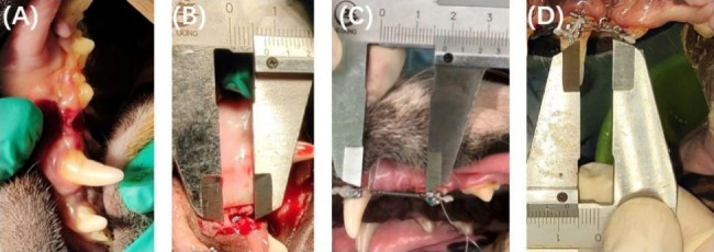
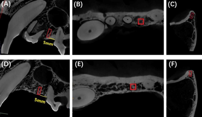
Methods: The bilateral maxillary first premolars of five beagle dogs were extracted and bone defects were created. The right and left sides of the maxilla were randomly divided into control and experimental sides. On the experimental side, the maxillary second premolar was mesially moved into the position of the missing first premolar. On the control side, the second maxillary premolar was extracted. Six months later, the beagles were euthanized. Microcomputer tomography was used to analyze bone microstructure parameters, alveolar bone height and alveolar bone width of the regenerated bone. Histological analysis was performed by staining tissue sections with toluidine blue.
Results: Median BV/TV values in the experimental group (81.78%) were significantly larger than those in the control group (35.67%; p = 0.04). Median Tb.Sp values in the experimental group (0.14 mm) were significantly lower than those in the control group (0.54 mm; p = 0.04). Median Tb.Th values in the experimental group (0.48 mm) were significantly higher than those in the control group (0.21 mm; p = 0.04). Median Tb.Pf values in the experimental group (0.65/mm) were significantly lower than those in the control group (3.15/mm; p = 0.04). There was no significant difference in the trabecular number (Tb.N) between the two groups (p = 0.23). The median alveolar bone height values in the experimental group (-0.81 mm) were significantly higher than those in the control group (-2.11 mm; p = 0.04) at a distance 5 mm from the mesial CEJ of the third premolar. The median alveolar bone height values in the experimental group (0.45 mm) were significantly higher than those in the control group (-1.70 mm; p = 0.04) at a distance 6 mm from the mesial CEJ of the third premolar. There was no significant difference in alveolar bone width when compared between the two groups (p > 0.05).
Conclusions: The newly formed edentulous area created by orthodontic treatment had more compact and thicker trabeculae than the extraction socket. Furthermore, the newly formed edentulous area had a greater alveolar bone height available for the placement of implants.
期刊介绍:
Head & Face Medicine is a multidisciplinary open access journal that publishes basic and clinical research concerning all aspects of cranial, facial and oral conditions.
The journal covers all aspects of cranial, facial and oral diseases and their management. It has been designed as a multidisciplinary journal for clinicians and researchers involved in the diagnostic and therapeutic aspects of diseases which affect the human head and face. The journal is wide-ranging, covering the development, aetiology, epidemiology and therapy of head and face diseases to the basic science that underlies these diseases. Management of head and face diseases includes all aspects of surgical and non-surgical treatments including psychopharmacological therapies.


 分享
分享
 求助内容:
求助内容: 应助结果提醒方式:
 扫码关注我们
