Dania G Malik, Tanya J Rath, Javier C Urcuyo Acevedo, Peter D Canoll, Kristin R Swanson, Jerrold L Boxerman, C Chad Quarles, Kathleen M Schmainda, Terry C Burns, Leland S Hu
{"title":"区分胶质瘤和治疗效果的先进 MRI 方案:技术现状与未来方向。","authors":"Dania G Malik, Tanya J Rath, Javier C Urcuyo Acevedo, Peter D Canoll, Kristin R Swanson, Jerrold L Boxerman, C Chad Quarles, Kathleen M Schmainda, Terry C Burns, Leland S Hu","doi":"10.3389/fradi.2022.809373","DOIUrl":null,"url":null,"abstract":"<p><p>In the follow-up treatment of high-grade gliomas (HGGs), differentiating true tumor progression from treatment-related effects, such as pseudoprogression and radiation necrosis, presents an ongoing clinical challenge. Conventional MRI with and without intravenous contrast serves as the clinical benchmark for the posttreatment surveillance imaging of HGG. However, many advanced imaging techniques have shown promise in helping better delineate the findings in indeterminate scenarios, as posttreatment effects can often mimic true tumor progression on conventional imaging. These challenges are further confounded by the histologic admixture that can commonly occur between tumor growth and treatment-related effects within the posttreatment bed. This review discusses the current practices in the surveillance imaging of HGG and the role of advanced imaging techniques, including perfusion MRI and metabolic MRI.</p>","PeriodicalId":73101,"journal":{"name":"Frontiers in radiology","volume":"2 ","pages":"809373"},"PeriodicalIF":2.3000,"publicationDate":"2022-04-15","publicationTypes":"Journal Article","fieldsOfStudy":null,"isOpenAccess":false,"openAccessPdf":"https://www.ncbi.nlm.nih.gov/pmc/articles/PMC10365126/pdf/","citationCount":"0","resultStr":"{\"title\":\"Advanced MRI Protocols to Discriminate Glioma From Treatment Effects: State of the Art and Future Directions.\",\"authors\":\"Dania G Malik, Tanya J Rath, Javier C Urcuyo Acevedo, Peter D Canoll, Kristin R Swanson, Jerrold L Boxerman, C Chad Quarles, Kathleen M Schmainda, Terry C Burns, Leland S Hu\",\"doi\":\"10.3389/fradi.2022.809373\",\"DOIUrl\":null,\"url\":null,\"abstract\":\"<p><p>In the follow-up treatment of high-grade gliomas (HGGs), differentiating true tumor progression from treatment-related effects, such as pseudoprogression and radiation necrosis, presents an ongoing clinical challenge. Conventional MRI with and without intravenous contrast serves as the clinical benchmark for the posttreatment surveillance imaging of HGG. However, many advanced imaging techniques have shown promise in helping better delineate the findings in indeterminate scenarios, as posttreatment effects can often mimic true tumor progression on conventional imaging. These challenges are further confounded by the histologic admixture that can commonly occur between tumor growth and treatment-related effects within the posttreatment bed. This review discusses the current practices in the surveillance imaging of HGG and the role of advanced imaging techniques, including perfusion MRI and metabolic MRI.</p>\",\"PeriodicalId\":73101,\"journal\":{\"name\":\"Frontiers in radiology\",\"volume\":\"2 \",\"pages\":\"809373\"},\"PeriodicalIF\":2.3000,\"publicationDate\":\"2022-04-15\",\"publicationTypes\":\"Journal Article\",\"fieldsOfStudy\":null,\"isOpenAccess\":false,\"openAccessPdf\":\"https://www.ncbi.nlm.nih.gov/pmc/articles/PMC10365126/pdf/\",\"citationCount\":\"0\",\"resultStr\":null,\"platform\":\"Semanticscholar\",\"paperid\":null,\"PeriodicalName\":\"Frontiers in radiology\",\"FirstCategoryId\":\"1085\",\"ListUrlMain\":\"https://doi.org/10.3389/fradi.2022.809373\",\"RegionNum\":0,\"RegionCategory\":null,\"ArticlePicture\":[],\"TitleCN\":null,\"AbstractTextCN\":null,\"PMCID\":null,\"EPubDate\":\"2022/1/1 0:00:00\",\"PubModel\":\"eCollection\",\"JCR\":\"\",\"JCRName\":\"\",\"Score\":null,\"Total\":0}","platform":"Semanticscholar","paperid":null,"PeriodicalName":"Frontiers in radiology","FirstCategoryId":"1085","ListUrlMain":"https://doi.org/10.3389/fradi.2022.809373","RegionNum":0,"RegionCategory":null,"ArticlePicture":[],"TitleCN":null,"AbstractTextCN":null,"PMCID":null,"EPubDate":"2022/1/1 0:00:00","PubModel":"eCollection","JCR":"","JCRName":"","Score":null,"Total":0}
Advanced MRI Protocols to Discriminate Glioma From Treatment Effects: State of the Art and Future Directions.
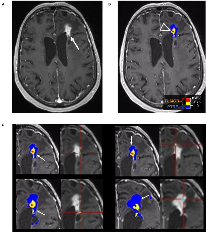
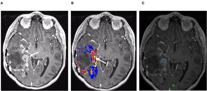
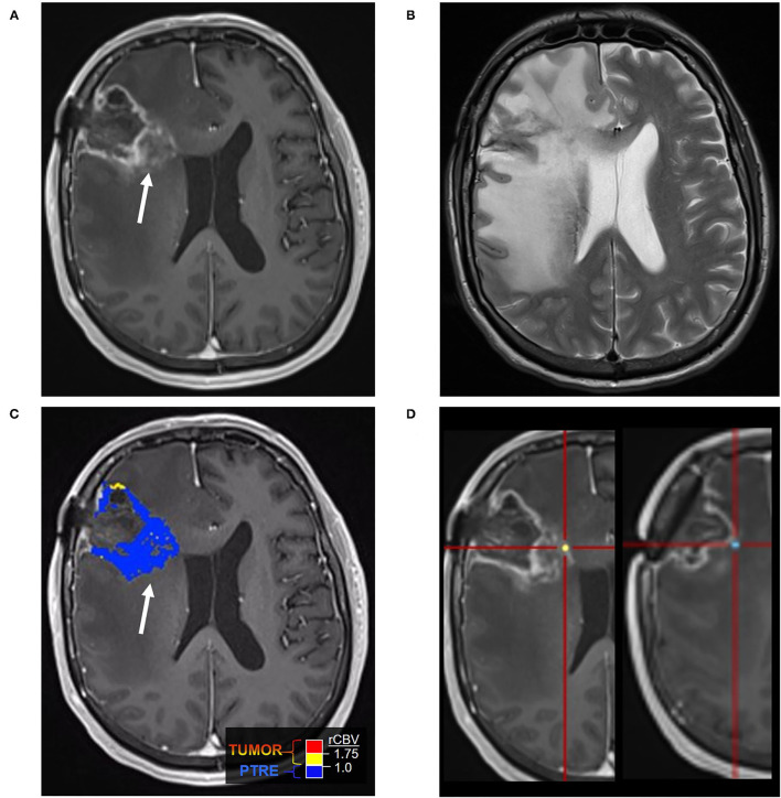
In the follow-up treatment of high-grade gliomas (HGGs), differentiating true tumor progression from treatment-related effects, such as pseudoprogression and radiation necrosis, presents an ongoing clinical challenge. Conventional MRI with and without intravenous contrast serves as the clinical benchmark for the posttreatment surveillance imaging of HGG. However, many advanced imaging techniques have shown promise in helping better delineate the findings in indeterminate scenarios, as posttreatment effects can often mimic true tumor progression on conventional imaging. These challenges are further confounded by the histologic admixture that can commonly occur between tumor growth and treatment-related effects within the posttreatment bed. This review discusses the current practices in the surveillance imaging of HGG and the role of advanced imaging techniques, including perfusion MRI and metabolic MRI.


 分享
分享

 求助内容:
求助内容: 应助结果提醒方式:
 扫码关注我们
