{"title":"阻生第三磨牙的位置可以作为病理状况的早期危险指标吗?回顾性锥束计算机断层扫描研究。","authors":"Melda Pelin Akkitap, Birsay Gumru","doi":"10.5037/jomr.2023.14203","DOIUrl":null,"url":null,"abstract":"<p><strong>Objectives: </strong>The aim of this retrospective study was to evaluate the prevalence of pathologies associated with impacted third molars in relation to tooth position on cone-beam computed tomography images.</p><p><strong>Material and methods: </strong>In 348 cone-beam computed tomography images, the position of 640 impacted third molars (mesiodistal angulation, buccolingual inclination, impaction depth, and contact point localization) and the presence of pathologies (distal caries, external root resorption, marginal bone loss, and pathological follicular space) were evaluated. The data were analysed statistically with a significance level set at P < 0.05.</p><p><strong>Results: </strong>Distal caries was mostly detected in relation to Class A (20.4%) and contact point at (12.5%) and above (10.5%) the cementoenamel junction (CEJ) (P = 0.000; P < 0.05). External root resorption and marginal bone loss were more common in mesioangular angulation (52.3% and 80.1%, respectively), Class C (53% and 73.8%, respectively), and contact point below the CEJ (53.2% and 73.3%, respectively) (P = 0.000; P < 0.05). Lingual inclination was identified as a new risk factor for associated pathologies (P < 0.05). Pathological follicular space was significantly more likely to occur in those with inverted angulation (100%) and absence of contact (31.5%) (P = 0.000 and P = 0.010, respectively; P < 0.05).</p><p><strong>Conclusions: </strong>Pathologies arising in second molars in relation to impacted third molars are significantly associated with the three-dimensional position of impacted third molars, and watchful monitoring or prophylactic removal of impacted third molars should be considered, taking into account the relevant risk parameters for the related pathologies.</p>","PeriodicalId":53254,"journal":{"name":"eJournal of Oral Maxillofacial Research","volume":"14 2","pages":"e3"},"PeriodicalIF":1.0000,"publicationDate":"2023-06-30","publicationTypes":"Journal Article","fieldsOfStudy":null,"isOpenAccess":false,"openAccessPdf":"https://ftp.ncbi.nlm.nih.gov/pub/pmc/oa_pdf/84/98/jomr-14-e3.PMC10382195.pdf","citationCount":"0","resultStr":"{\"title\":\"Can the Position of the Impacted Third Molars Be an Early Risk Indicator of Pathological Conditions? A Retrospective Cone-Beam Computed Tomography Study.\",\"authors\":\"Melda Pelin Akkitap, Birsay Gumru\",\"doi\":\"10.5037/jomr.2023.14203\",\"DOIUrl\":null,\"url\":null,\"abstract\":\"<p><strong>Objectives: </strong>The aim of this retrospective study was to evaluate the prevalence of pathologies associated with impacted third molars in relation to tooth position on cone-beam computed tomography images.</p><p><strong>Material and methods: </strong>In 348 cone-beam computed tomography images, the position of 640 impacted third molars (mesiodistal angulation, buccolingual inclination, impaction depth, and contact point localization) and the presence of pathologies (distal caries, external root resorption, marginal bone loss, and pathological follicular space) were evaluated. The data were analysed statistically with a significance level set at P < 0.05.</p><p><strong>Results: </strong>Distal caries was mostly detected in relation to Class A (20.4%) and contact point at (12.5%) and above (10.5%) the cementoenamel junction (CEJ) (P = 0.000; P < 0.05). External root resorption and marginal bone loss were more common in mesioangular angulation (52.3% and 80.1%, respectively), Class C (53% and 73.8%, respectively), and contact point below the CEJ (53.2% and 73.3%, respectively) (P = 0.000; P < 0.05). Lingual inclination was identified as a new risk factor for associated pathologies (P < 0.05). Pathological follicular space was significantly more likely to occur in those with inverted angulation (100%) and absence of contact (31.5%) (P = 0.000 and P = 0.010, respectively; P < 0.05).</p><p><strong>Conclusions: </strong>Pathologies arising in second molars in relation to impacted third molars are significantly associated with the three-dimensional position of impacted third molars, and watchful monitoring or prophylactic removal of impacted third molars should be considered, taking into account the relevant risk parameters for the related pathologies.</p>\",\"PeriodicalId\":53254,\"journal\":{\"name\":\"eJournal of Oral Maxillofacial Research\",\"volume\":\"14 2\",\"pages\":\"e3\"},\"PeriodicalIF\":1.0000,\"publicationDate\":\"2023-06-30\",\"publicationTypes\":\"Journal Article\",\"fieldsOfStudy\":null,\"isOpenAccess\":false,\"openAccessPdf\":\"https://ftp.ncbi.nlm.nih.gov/pub/pmc/oa_pdf/84/98/jomr-14-e3.PMC10382195.pdf\",\"citationCount\":\"0\",\"resultStr\":null,\"platform\":\"Semanticscholar\",\"paperid\":null,\"PeriodicalName\":\"eJournal of Oral Maxillofacial Research\",\"FirstCategoryId\":\"1085\",\"ListUrlMain\":\"https://doi.org/10.5037/jomr.2023.14203\",\"RegionNum\":0,\"RegionCategory\":null,\"ArticlePicture\":[],\"TitleCN\":null,\"AbstractTextCN\":null,\"PMCID\":null,\"EPubDate\":\"2023/4/1 0:00:00\",\"PubModel\":\"eCollection\",\"JCR\":\"Q3\",\"JCRName\":\"DENTISTRY, ORAL SURGERY & MEDICINE\",\"Score\":null,\"Total\":0}","platform":"Semanticscholar","paperid":null,"PeriodicalName":"eJournal of Oral Maxillofacial Research","FirstCategoryId":"1085","ListUrlMain":"https://doi.org/10.5037/jomr.2023.14203","RegionNum":0,"RegionCategory":null,"ArticlePicture":[],"TitleCN":null,"AbstractTextCN":null,"PMCID":null,"EPubDate":"2023/4/1 0:00:00","PubModel":"eCollection","JCR":"Q3","JCRName":"DENTISTRY, ORAL SURGERY & MEDICINE","Score":null,"Total":0}
引用次数: 0
摘要
目的:本回顾性研究的目的是评估与锥束计算机断层扫描图像上牙齿位置有关的第三磨牙阻生病理的患病率。材料和方法:在348张锥形束计算机断层图像中,评估640颗阻生第三磨牙的位置(中远端角度、颊舌倾斜、嵌塞深度和接触点定位)和病理(远端龋齿、外根吸收、边缘骨丢失和病理性滤泡间隙)的存在。对数据进行统计学分析,P < 0.05为显著水平。结果:远端龋多见于A类(20.4%)和接触点(12.5%)及以上(10.5%)牙髓-牙釉质交界处(CEJ) (P = 0.000; P < 0.05)。外根吸收和边缘骨丢失在中角角度(分别为52.3%和80.1%)、C类(分别为53%和73.8%)和CEJ以下接触点(分别为53.2%和73.3%)较为常见(P = 0.000; P < 0.05)。舌倾斜被确定为新的相关病理危险因素(P < 0.05)。病理性滤泡间隙的发生在倒角组(100%)和无接触组(31.5%)(P = 0.000和P = 0.010, P < 0.05)。结论:阻生第三磨牙发生的第二磨牙病变与阻生第三磨牙的三维位置密切相关,应考虑相关病变的相关风险参数,观察监测或预防性拔除阻生第三磨牙。
Can the Position of the Impacted Third Molars Be an Early Risk Indicator of Pathological Conditions? A Retrospective Cone-Beam Computed Tomography Study.
Objectives: The aim of this retrospective study was to evaluate the prevalence of pathologies associated with impacted third molars in relation to tooth position on cone-beam computed tomography images.
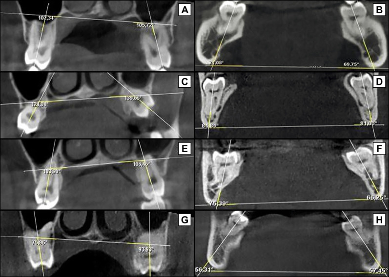
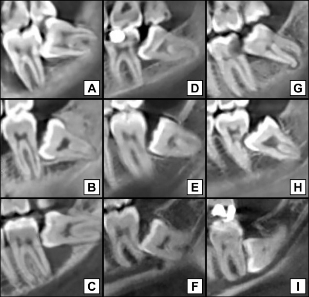
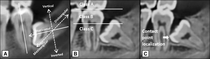
Material and methods: In 348 cone-beam computed tomography images, the position of 640 impacted third molars (mesiodistal angulation, buccolingual inclination, impaction depth, and contact point localization) and the presence of pathologies (distal caries, external root resorption, marginal bone loss, and pathological follicular space) were evaluated. The data were analysed statistically with a significance level set at P < 0.05.
Results: Distal caries was mostly detected in relation to Class A (20.4%) and contact point at (12.5%) and above (10.5%) the cementoenamel junction (CEJ) (P = 0.000; P < 0.05). External root resorption and marginal bone loss were more common in mesioangular angulation (52.3% and 80.1%, respectively), Class C (53% and 73.8%, respectively), and contact point below the CEJ (53.2% and 73.3%, respectively) (P = 0.000; P < 0.05). Lingual inclination was identified as a new risk factor for associated pathologies (P < 0.05). Pathological follicular space was significantly more likely to occur in those with inverted angulation (100%) and absence of contact (31.5%) (P = 0.000 and P = 0.010, respectively; P < 0.05).
Conclusions: Pathologies arising in second molars in relation to impacted third molars are significantly associated with the three-dimensional position of impacted third molars, and watchful monitoring or prophylactic removal of impacted third molars should be considered, taking into account the relevant risk parameters for the related pathologies.


 分享
分享

 求助内容:
求助内容: 应助结果提醒方式:
 扫码关注我们
