{"title":"用于原位光学分析的标准96孔基高通量微流体灌注生物膜反应器。","authors":"David McLeod, Lai Wei, Zhenyu Li","doi":"10.1007/s10544-023-00668-w","DOIUrl":null,"url":null,"abstract":"<div><p>Biofilm infections represent a major public health threat due to their high tolerance to antimicrobials and the lack of specific anti-biofilm drugs. To develop such drugs, it is crucial to have high-throughput biofilm growth systems that can emulate <i>in vivo</i> conditions without the cost and complexity of animal models. However, no current biofilm reactor can provide <i>in vivo</i>-like conditions in a high throughput standard microtiter format. This paper demonstrates a novel high-throughput (HT) microfluidic perfusion biofilm reactor (HT-μPBR) compatible with a standard 96-well microtiter plate for <i>in situ</i> optical analysis. A snap-on liquid-tight cover for standard microtiter plates was designed and fabricated with fluidic channels to provide closed-loop recirculating perfusion. Our system takes steps toward providing <i>in vivo</i>-like conditions with controlled shear stress and nutrient delivery. We describe the system fabrication and usage in optical analysis of biomass and viability of <i>Escherichia coli</i> (<i>E. coli</i>) biofilms. The HT-μPBR was set to perfuse at 1 mL/min corresponding to an average shear rate of approximately <span>\\(5.7{\\mathrm{s}}^{-1}\\)</span> on the bottom surface of a single well. Biofilms were detected on well plate bottoms and measured using a fluorescence microscope and plate reader to determine biomass and viability. Samples cultured in the HT-μPBR showed increased biomass while maintaining viability after 24 h. The HT-μPBR can further be combined with HT antibiotic susceptibility testing and additional optical techniques such as time-lapse imaging to improve understanding of the drug reaction mechanism as well as the optimization of drug combinations and delivery profiles.</p></div>","PeriodicalId":490,"journal":{"name":"Biomedical Microdevices","volume":"25 3","pages":""},"PeriodicalIF":3.3000,"publicationDate":"2023-07-26","publicationTypes":"Journal Article","fieldsOfStudy":null,"isOpenAccess":false,"openAccessPdf":"","citationCount":"0","resultStr":"{\"title\":\"A standard 96-well based high throughput microfluidic perfusion biofilm reactor for in situ optical analysis\",\"authors\":\"David McLeod, Lai Wei, Zhenyu Li\",\"doi\":\"10.1007/s10544-023-00668-w\",\"DOIUrl\":null,\"url\":null,\"abstract\":\"<div><p>Biofilm infections represent a major public health threat due to their high tolerance to antimicrobials and the lack of specific anti-biofilm drugs. To develop such drugs, it is crucial to have high-throughput biofilm growth systems that can emulate <i>in vivo</i> conditions without the cost and complexity of animal models. However, no current biofilm reactor can provide <i>in vivo</i>-like conditions in a high throughput standard microtiter format. This paper demonstrates a novel high-throughput (HT) microfluidic perfusion biofilm reactor (HT-μPBR) compatible with a standard 96-well microtiter plate for <i>in situ</i> optical analysis. A snap-on liquid-tight cover for standard microtiter plates was designed and fabricated with fluidic channels to provide closed-loop recirculating perfusion. Our system takes steps toward providing <i>in vivo</i>-like conditions with controlled shear stress and nutrient delivery. We describe the system fabrication and usage in optical analysis of biomass and viability of <i>Escherichia coli</i> (<i>E. coli</i>) biofilms. The HT-μPBR was set to perfuse at 1 mL/min corresponding to an average shear rate of approximately <span>\\\\(5.7{\\\\mathrm{s}}^{-1}\\\\)</span> on the bottom surface of a single well. Biofilms were detected on well plate bottoms and measured using a fluorescence microscope and plate reader to determine biomass and viability. Samples cultured in the HT-μPBR showed increased biomass while maintaining viability after 24 h. The HT-μPBR can further be combined with HT antibiotic susceptibility testing and additional optical techniques such as time-lapse imaging to improve understanding of the drug reaction mechanism as well as the optimization of drug combinations and delivery profiles.</p></div>\",\"PeriodicalId\":490,\"journal\":{\"name\":\"Biomedical Microdevices\",\"volume\":\"25 3\",\"pages\":\"\"},\"PeriodicalIF\":3.3000,\"publicationDate\":\"2023-07-26\",\"publicationTypes\":\"Journal Article\",\"fieldsOfStudy\":null,\"isOpenAccess\":false,\"openAccessPdf\":\"\",\"citationCount\":\"0\",\"resultStr\":null,\"platform\":\"Semanticscholar\",\"paperid\":null,\"PeriodicalName\":\"Biomedical Microdevices\",\"FirstCategoryId\":\"5\",\"ListUrlMain\":\"https://link.springer.com/article/10.1007/s10544-023-00668-w\",\"RegionNum\":4,\"RegionCategory\":\"医学\",\"ArticlePicture\":[],\"TitleCN\":null,\"AbstractTextCN\":null,\"PMCID\":null,\"EPubDate\":\"\",\"PubModel\":\"\",\"JCR\":\"Q3\",\"JCRName\":\"ENGINEERING, BIOMEDICAL\",\"Score\":null,\"Total\":0}","platform":"Semanticscholar","paperid":null,"PeriodicalName":"Biomedical Microdevices","FirstCategoryId":"5","ListUrlMain":"https://link.springer.com/article/10.1007/s10544-023-00668-w","RegionNum":4,"RegionCategory":"医学","ArticlePicture":[],"TitleCN":null,"AbstractTextCN":null,"PMCID":null,"EPubDate":"","PubModel":"","JCR":"Q3","JCRName":"ENGINEERING, BIOMEDICAL","Score":null,"Total":0}
A standard 96-well based high throughput microfluidic perfusion biofilm reactor for in situ optical analysis
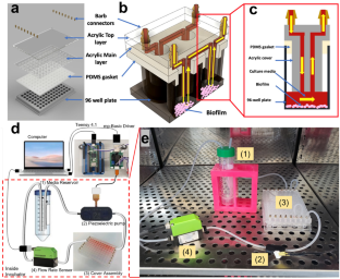
Biofilm infections represent a major public health threat due to their high tolerance to antimicrobials and the lack of specific anti-biofilm drugs. To develop such drugs, it is crucial to have high-throughput biofilm growth systems that can emulate in vivo conditions without the cost and complexity of animal models. However, no current biofilm reactor can provide in vivo-like conditions in a high throughput standard microtiter format. This paper demonstrates a novel high-throughput (HT) microfluidic perfusion biofilm reactor (HT-μPBR) compatible with a standard 96-well microtiter plate for in situ optical analysis. A snap-on liquid-tight cover for standard microtiter plates was designed and fabricated with fluidic channels to provide closed-loop recirculating perfusion. Our system takes steps toward providing in vivo-like conditions with controlled shear stress and nutrient delivery. We describe the system fabrication and usage in optical analysis of biomass and viability of Escherichia coli (E. coli) biofilms. The HT-μPBR was set to perfuse at 1 mL/min corresponding to an average shear rate of approximately \(5.7{\mathrm{s}}^{-1}\) on the bottom surface of a single well. Biofilms were detected on well plate bottoms and measured using a fluorescence microscope and plate reader to determine biomass and viability. Samples cultured in the HT-μPBR showed increased biomass while maintaining viability after 24 h. The HT-μPBR can further be combined with HT antibiotic susceptibility testing and additional optical techniques such as time-lapse imaging to improve understanding of the drug reaction mechanism as well as the optimization of drug combinations and delivery profiles.
期刊介绍:
Biomedical Microdevices: BioMEMS and Biomedical Nanotechnology is an interdisciplinary periodical devoted to all aspects of research in the medical diagnostic and therapeutic applications of Micro-Electro-Mechanical Systems (BioMEMS) and nanotechnology for medicine and biology.
General subjects of interest include the design, characterization, testing, modeling and clinical validation of microfabricated systems, and their integration on-chip and in larger functional units. The specific interests of the Journal include systems for neural stimulation and recording, bioseparation technologies such as nanofilters and electrophoretic equipment, miniaturized analytic and DNA identification systems, biosensors, and micro/nanotechnologies for cell and tissue research, tissue engineering, cell transplantation, and the controlled release of drugs and biological molecules.
Contributions reporting on fundamental and applied investigations of the material science, biochemistry, and physics of biomedical microdevices and nanotechnology are encouraged. A non-exhaustive list of fields of interest includes: nanoparticle synthesis, characterization, and validation of therapeutic or imaging efficacy in animal models; biocompatibility; biochemical modification of microfabricated devices, with reference to non-specific protein adsorption, and the active immobilization and patterning of proteins on micro/nanofabricated surfaces; the dynamics of fluids in micro-and-nano-fabricated channels; the electromechanical and structural response of micro/nanofabricated systems; the interactions of microdevices with cells and tissues, including biocompatibility and biodegradation studies; variations in the characteristics of the systems as a function of the micro/nanofabrication parameters.


 分享
分享
 求助内容:
求助内容: 应助结果提醒方式:
 扫码关注我们
