{"title":"次氯酸钠浓度对人血凝块纤维蛋白结构和转化生长因子- β 1释放的影响:体外研究","authors":"Anisha Mishra, Velmurugan Natanasabapathy, Nandini Suresh","doi":"10.5395/rde.2022.47.e42","DOIUrl":null,"url":null,"abstract":"<p><strong>Objective: </strong>This study investigated the effects of various concentrations of sodium hypochlorite (NaOCl) on human whole-blood clotting kinetics, the structure of the blood clots formed, and transforming growth factor (TGF)-β1 release.</p><p><strong>Materials and methods: </strong>Human whole blood was collected from 5 healthy volunteers and divided into 4 groups: CG (control, 0.5 mL of blood), BN<sub>0.5</sub> (0.5 mL of blood with 0.5 mL of 0.5% NaOCl), BN<sub>3</sub> (0.5 mL of blood with 0.5 mL of 3% NaOCl), and BN<sub>5.25</sub> (0.5 mL of blood with 0.5 mL of 5.25% NaOCl). The effects of NaOCl on clotting kinetics, structure of fibrin and cells, and release of TGF-β1 were assessed using thromboelastography (TEG), scanning electron microscopy (SEM), and enzyme-linked immunosobent assay, respectively. Statistical analysis was conducted using the Kruskal Wallis and Mann-Whitney <i>U</i> tests, followed by the <i>post hoc</i> Dunn test. A <i>p</i> value < 0.05 indicated statistical significance.</p><p><strong>Results: </strong>The blood samples in BN<sub>0.5</sub> and BN<sub>3</sub> did not clot, whereas the TEG of BN<sub>5.25</sub> showed altered clot formation. Samples from the CG and BN<sub>3</sub> groups could only be processed with SEM, which showed that the latter lacked fibrin formation and branching of fibers, as well as clumping of red blood cells with surface roughening and distortion. TGF-β1 release was significantly highest in BN<sub>3</sub> when all groups were compared to CG (<i>p</i> < 0.05).</p><p><strong>Conclusions: </strong>Each concentration of NaOCl affected the release of TGF-β1 from blood clots and altered the clotting mechanism of blood by affecting clotting kinetics and cell structure.</p>","PeriodicalId":21102,"journal":{"name":"Restorative Dentistry & Endodontics","volume":"47 4","pages":"e42"},"PeriodicalIF":0.0000,"publicationDate":"2022-11-01","publicationTypes":"Journal Article","fieldsOfStudy":null,"isOpenAccess":false,"openAccessPdf":"https://ftp.ncbi.nlm.nih.gov/pub/pmc/oa_pdf/e0/94/rde-47-e42.PMC9715373.pdf","citationCount":"0","resultStr":"{\"title\":\"The influence of sodium hypochlorite concentration on the fibrin structure of human blood clots and transforming growth factor-beta 1 release: an <i>ex vivo</i> study.\",\"authors\":\"Anisha Mishra, Velmurugan Natanasabapathy, Nandini Suresh\",\"doi\":\"10.5395/rde.2022.47.e42\",\"DOIUrl\":null,\"url\":null,\"abstract\":\"<p><strong>Objective: </strong>This study investigated the effects of various concentrations of sodium hypochlorite (NaOCl) on human whole-blood clotting kinetics, the structure of the blood clots formed, and transforming growth factor (TGF)-β1 release.</p><p><strong>Materials and methods: </strong>Human whole blood was collected from 5 healthy volunteers and divided into 4 groups: CG (control, 0.5 mL of blood), BN<sub>0.5</sub> (0.5 mL of blood with 0.5 mL of 0.5% NaOCl), BN<sub>3</sub> (0.5 mL of blood with 0.5 mL of 3% NaOCl), and BN<sub>5.25</sub> (0.5 mL of blood with 0.5 mL of 5.25% NaOCl). The effects of NaOCl on clotting kinetics, structure of fibrin and cells, and release of TGF-β1 were assessed using thromboelastography (TEG), scanning electron microscopy (SEM), and enzyme-linked immunosobent assay, respectively. Statistical analysis was conducted using the Kruskal Wallis and Mann-Whitney <i>U</i> tests, followed by the <i>post hoc</i> Dunn test. A <i>p</i> value < 0.05 indicated statistical significance.</p><p><strong>Results: </strong>The blood samples in BN<sub>0.5</sub> and BN<sub>3</sub> did not clot, whereas the TEG of BN<sub>5.25</sub> showed altered clot formation. Samples from the CG and BN<sub>3</sub> groups could only be processed with SEM, which showed that the latter lacked fibrin formation and branching of fibers, as well as clumping of red blood cells with surface roughening and distortion. TGF-β1 release was significantly highest in BN<sub>3</sub> when all groups were compared to CG (<i>p</i> < 0.05).</p><p><strong>Conclusions: </strong>Each concentration of NaOCl affected the release of TGF-β1 from blood clots and altered the clotting mechanism of blood by affecting clotting kinetics and cell structure.</p>\",\"PeriodicalId\":21102,\"journal\":{\"name\":\"Restorative Dentistry & Endodontics\",\"volume\":\"47 4\",\"pages\":\"e42\"},\"PeriodicalIF\":0.0000,\"publicationDate\":\"2022-11-01\",\"publicationTypes\":\"Journal Article\",\"fieldsOfStudy\":null,\"isOpenAccess\":false,\"openAccessPdf\":\"https://ftp.ncbi.nlm.nih.gov/pub/pmc/oa_pdf/e0/94/rde-47-e42.PMC9715373.pdf\",\"citationCount\":\"0\",\"resultStr\":null,\"platform\":\"Semanticscholar\",\"paperid\":null,\"PeriodicalName\":\"Restorative Dentistry & Endodontics\",\"FirstCategoryId\":\"1085\",\"ListUrlMain\":\"https://doi.org/10.5395/rde.2022.47.e42\",\"RegionNum\":0,\"RegionCategory\":null,\"ArticlePicture\":[],\"TitleCN\":null,\"AbstractTextCN\":null,\"PMCID\":null,\"EPubDate\":\"\",\"PubModel\":\"\",\"JCR\":\"\",\"JCRName\":\"\",\"Score\":null,\"Total\":0}","platform":"Semanticscholar","paperid":null,"PeriodicalName":"Restorative Dentistry & Endodontics","FirstCategoryId":"1085","ListUrlMain":"https://doi.org/10.5395/rde.2022.47.e42","RegionNum":0,"RegionCategory":null,"ArticlePicture":[],"TitleCN":null,"AbstractTextCN":null,"PMCID":null,"EPubDate":"","PubModel":"","JCR":"","JCRName":"","Score":null,"Total":0}
引用次数: 0
摘要
目的:研究不同浓度的次氯酸钠(NaOCl)对人全血凝血动力学、形成的血凝块结构及转化生长因子(TGF)-β1释放的影响。材料与方法:采集5名健康志愿者的人全血,分为4组:CG(对照组,0.5 mL血)、BN0.5 (0.5 mL血,0.5 mL 0.5% NaOCl)、BN3 (0.5 mL血,0.5 mL 3% NaOCl)、BN5.25 (0.5 mL血,0.5 mL 5.25% NaOCl)。分别采用血栓弹性成像(TEG)、扫描电镜(SEM)和酶联免疫吸附法评估NaOCl对凝血动力学、纤维蛋白和细胞结构以及TGF-β1释放的影响。采用Kruskal Wallis检验和Mann-Whitney U检验进行统计分析,随后采用事后Dunn检验。p值< 0.05为有统计学意义。结果:BN0.5和BN3的血样品未形成凝块,而BN5.25的TEG出现了凝块形成的改变。CG组和BN3组的样品只能通过扫描电镜进行处理,发现后者缺乏纤维蛋白形成和纤维分支,红细胞团块,表面粗糙和扭曲。与CG比较,BN3组TGF-β1释放量最高(p < 0.05)。结论:不同浓度的NaOCl通过影响凝血动力学和细胞结构,影响凝血块中TGF-β1的释放,改变血液的凝血机制。
The influence of sodium hypochlorite concentration on the fibrin structure of human blood clots and transforming growth factor-beta 1 release: an ex vivo study.
Objective: This study investigated the effects of various concentrations of sodium hypochlorite (NaOCl) on human whole-blood clotting kinetics, the structure of the blood clots formed, and transforming growth factor (TGF)-β1 release.
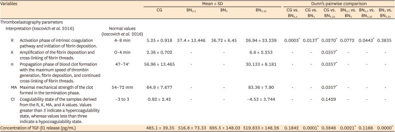
Materials and methods: Human whole blood was collected from 5 healthy volunteers and divided into 4 groups: CG (control, 0.5 mL of blood), BN0.5 (0.5 mL of blood with 0.5 mL of 0.5% NaOCl), BN3 (0.5 mL of blood with 0.5 mL of 3% NaOCl), and BN5.25 (0.5 mL of blood with 0.5 mL of 5.25% NaOCl). The effects of NaOCl on clotting kinetics, structure of fibrin and cells, and release of TGF-β1 were assessed using thromboelastography (TEG), scanning electron microscopy (SEM), and enzyme-linked immunosobent assay, respectively. Statistical analysis was conducted using the Kruskal Wallis and Mann-Whitney U tests, followed by the post hoc Dunn test. A p value < 0.05 indicated statistical significance.
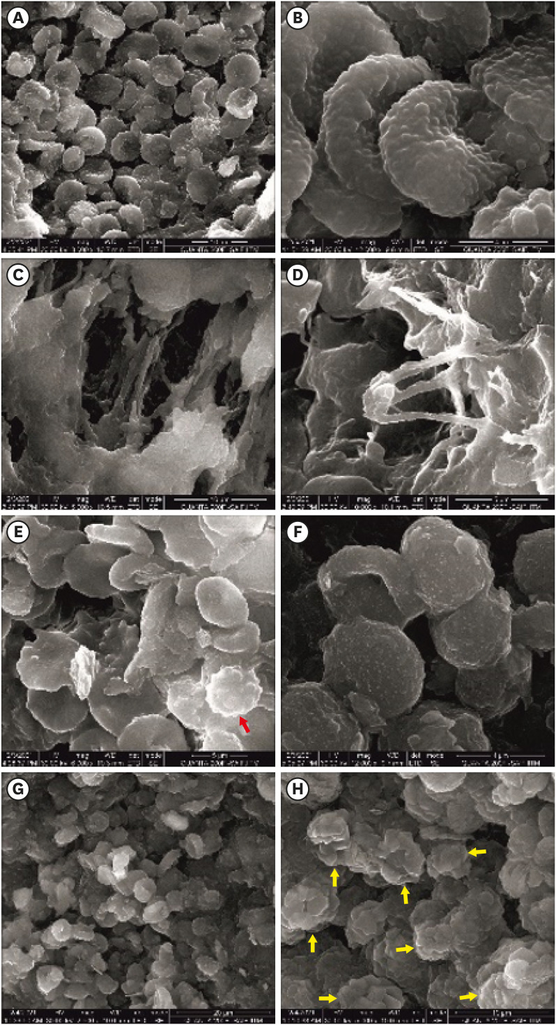
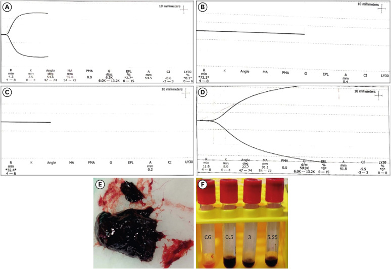
Results: The blood samples in BN0.5 and BN3 did not clot, whereas the TEG of BN5.25 showed altered clot formation. Samples from the CG and BN3 groups could only be processed with SEM, which showed that the latter lacked fibrin formation and branching of fibers, as well as clumping of red blood cells with surface roughening and distortion. TGF-β1 release was significantly highest in BN3 when all groups were compared to CG (p < 0.05).
Conclusions: Each concentration of NaOCl affected the release of TGF-β1 from blood clots and altered the clotting mechanism of blood by affecting clotting kinetics and cell structure.


 分享
分享

 求助内容:
求助内容: 应助结果提醒方式:
 扫码关注我们
