{"title":"在自然感染牛白血病病毒(BLV)的牛疾病进展过程中,携带BLV整合位点的细胞大量耗损后克隆扩增的可视化:一例报告","authors":"Susumu Saito, Kazuyoshi Hosomichi, Meripet Polat Yamanaka, Tetsuya Mizutani, Shin-Nosuke Takeshima, Yoko Aida","doi":"10.1186/s12977-022-00609-0","DOIUrl":null,"url":null,"abstract":"<p><p>Bovine leukemia virus (BLV) infects cattle, integrates into host DNA as a provirus, and induces malignant B-cell lymphoma. Previous studies have addressed the impact of proviral integration of BLV on BLV-induced leukemogenesis. However, no studies have monitored sequential changes in integration sites in which naturally infected BLV individuals progress from the premalignant stage to the terminal disease. Here, we collected blood samples from a single, naturally infected Holstein cow at three disease progression stages (Stage I: polyclonal stage, Stage II: polyclonal toward oligoclonal stage, Stage III: oligoclonal stage) and successfully visualized the kinetics of clonal expansion of cells carrying BLV integration sites using our BLV proviral DNA-capture sequencing method. Although 24 integration sites were detected in Stages I and II, 92% of these sites experienced massive depletion in Stage III. Of these sites, 46%, 37%, and 17% were located within introns of Refseq genes, intergenic regions, and repetitive sequences, respectively. At Stage III cattle with lymphoma, only two integration sites were generated de novo in the intergenic region of Chr1, and the intron of the CHEK2 gene on Chr17 was significantly increased. Our results are the first to demonstrate clonal expansion after the massive depletion of cells carrying BLV integration sites in a naturally infected cow.</p>","PeriodicalId":21123,"journal":{"name":"Retrovirology","volume":"19 1","pages":"24"},"PeriodicalIF":2.7000,"publicationDate":"2022-11-03","publicationTypes":"Journal Article","fieldsOfStudy":null,"isOpenAccess":false,"openAccessPdf":"https://www.ncbi.nlm.nih.gov/pmc/articles/PMC9635170/pdf/","citationCount":"1","resultStr":"{\"title\":\"Visualization of clonal expansion after massive depletion of cells carrying the bovine leukemia virus (BLV) integration sites during the course of disease progression in a BLV naturally-infected cow: a case report.\",\"authors\":\"Susumu Saito, Kazuyoshi Hosomichi, Meripet Polat Yamanaka, Tetsuya Mizutani, Shin-Nosuke Takeshima, Yoko Aida\",\"doi\":\"10.1186/s12977-022-00609-0\",\"DOIUrl\":null,\"url\":null,\"abstract\":\"<p><p>Bovine leukemia virus (BLV) infects cattle, integrates into host DNA as a provirus, and induces malignant B-cell lymphoma. Previous studies have addressed the impact of proviral integration of BLV on BLV-induced leukemogenesis. However, no studies have monitored sequential changes in integration sites in which naturally infected BLV individuals progress from the premalignant stage to the terminal disease. Here, we collected blood samples from a single, naturally infected Holstein cow at three disease progression stages (Stage I: polyclonal stage, Stage II: polyclonal toward oligoclonal stage, Stage III: oligoclonal stage) and successfully visualized the kinetics of clonal expansion of cells carrying BLV integration sites using our BLV proviral DNA-capture sequencing method. Although 24 integration sites were detected in Stages I and II, 92% of these sites experienced massive depletion in Stage III. Of these sites, 46%, 37%, and 17% were located within introns of Refseq genes, intergenic regions, and repetitive sequences, respectively. At Stage III cattle with lymphoma, only two integration sites were generated de novo in the intergenic region of Chr1, and the intron of the CHEK2 gene on Chr17 was significantly increased. Our results are the first to demonstrate clonal expansion after the massive depletion of cells carrying BLV integration sites in a naturally infected cow.</p>\",\"PeriodicalId\":21123,\"journal\":{\"name\":\"Retrovirology\",\"volume\":\"19 1\",\"pages\":\"24\"},\"PeriodicalIF\":2.7000,\"publicationDate\":\"2022-11-03\",\"publicationTypes\":\"Journal Article\",\"fieldsOfStudy\":null,\"isOpenAccess\":false,\"openAccessPdf\":\"https://www.ncbi.nlm.nih.gov/pmc/articles/PMC9635170/pdf/\",\"citationCount\":\"1\",\"resultStr\":null,\"platform\":\"Semanticscholar\",\"paperid\":null,\"PeriodicalName\":\"Retrovirology\",\"FirstCategoryId\":\"3\",\"ListUrlMain\":\"https://doi.org/10.1186/s12977-022-00609-0\",\"RegionNum\":3,\"RegionCategory\":\"医学\",\"ArticlePicture\":[],\"TitleCN\":null,\"AbstractTextCN\":null,\"PMCID\":null,\"EPubDate\":\"\",\"PubModel\":\"\",\"JCR\":\"Q3\",\"JCRName\":\"VIROLOGY\",\"Score\":null,\"Total\":0}","platform":"Semanticscholar","paperid":null,"PeriodicalName":"Retrovirology","FirstCategoryId":"3","ListUrlMain":"https://doi.org/10.1186/s12977-022-00609-0","RegionNum":3,"RegionCategory":"医学","ArticlePicture":[],"TitleCN":null,"AbstractTextCN":null,"PMCID":null,"EPubDate":"","PubModel":"","JCR":"Q3","JCRName":"VIROLOGY","Score":null,"Total":0}
Visualization of clonal expansion after massive depletion of cells carrying the bovine leukemia virus (BLV) integration sites during the course of disease progression in a BLV naturally-infected cow: a case report.
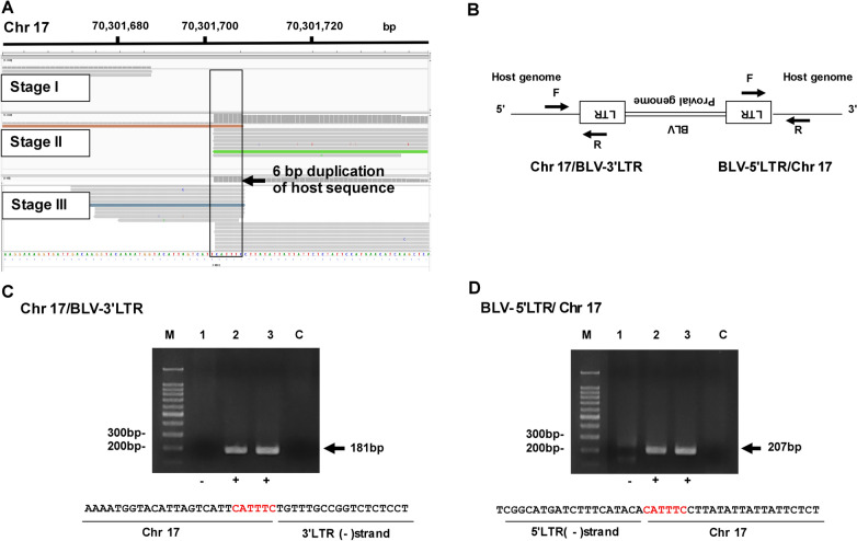
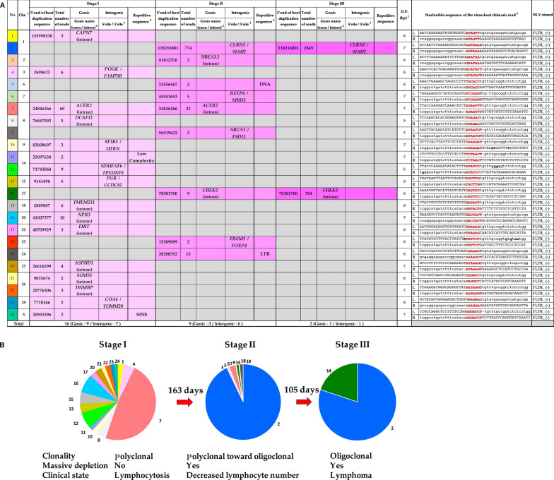
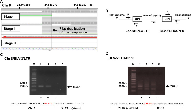
Bovine leukemia virus (BLV) infects cattle, integrates into host DNA as a provirus, and induces malignant B-cell lymphoma. Previous studies have addressed the impact of proviral integration of BLV on BLV-induced leukemogenesis. However, no studies have monitored sequential changes in integration sites in which naturally infected BLV individuals progress from the premalignant stage to the terminal disease. Here, we collected blood samples from a single, naturally infected Holstein cow at three disease progression stages (Stage I: polyclonal stage, Stage II: polyclonal toward oligoclonal stage, Stage III: oligoclonal stage) and successfully visualized the kinetics of clonal expansion of cells carrying BLV integration sites using our BLV proviral DNA-capture sequencing method. Although 24 integration sites were detected in Stages I and II, 92% of these sites experienced massive depletion in Stage III. Of these sites, 46%, 37%, and 17% were located within introns of Refseq genes, intergenic regions, and repetitive sequences, respectively. At Stage III cattle with lymphoma, only two integration sites were generated de novo in the intergenic region of Chr1, and the intron of the CHEK2 gene on Chr17 was significantly increased. Our results are the first to demonstrate clonal expansion after the massive depletion of cells carrying BLV integration sites in a naturally infected cow.
期刊介绍:
Retrovirology is an open access, online journal that publishes stringently peer-reviewed, high-impact articles on host-pathogen interactions, fundamental mechanisms of replication, immune defenses, animal models, and clinical science relating to retroviruses. Retroviruses are pleiotropically found in animals. Well-described examples include avian, murine and primate retroviruses.
Two human retroviruses are especially important pathogens. These are the human immunodeficiency virus, HIV, and the human T-cell leukemia virus, HTLV. HIV causes AIDS while HTLV-1 is the etiological agent for adult T-cell leukemia and HTLV-1-associated myelopathy/tropical spastic paraparesis. Retrovirology aims to cover comprehensively all aspects of human and animal retrovirus research.


 分享
分享
 求助内容:
求助内容: 应助结果提醒方式:
 扫码关注我们
