Xin Zhong, Tao-Tao Tang, An-Ran Shen, Jing-Yuan Cao, Jing Jing, Cui Wang, Xiao-Xiao Zhu, Yi Wen, Zuo-Lin Li, Bin Wang, Suo-Fu Qin, Bi-Cheng Liu, Lin-Li Lv
{"title":"小管上皮细胞来源的细胞外小泡vegf - a促进缺血性肾损伤的小管周围毛细血管修复。","authors":"Xin Zhong, Tao-Tao Tang, An-Ran Shen, Jing-Yuan Cao, Jing Jing, Cui Wang, Xiao-Xiao Zhu, Yi Wen, Zuo-Lin Li, Bin Wang, Suo-Fu Qin, Bi-Cheng Liu, Lin-Li Lv","doi":"10.1038/s41536-022-00268-x","DOIUrl":null,"url":null,"abstract":"<p><p>Peritubular capillaries (PTCs) are closely related to renal tubules in structure and function, and both are pivotal regulators in the development and progression of acute kidney injury (AKI). However, the mechanisms that underlie the interaction between PTCs and tubules during AKI remain unclear. Here we explored a new mode of tubulovascular crosstalk mediated by small extracellular vesicles (sEV) after AKI. In response to renal ischemia/reperfusion (I/R) injury, endothelial proliferation of PTCs and tubular expression of vascular endothelial growth factor-A (VEGF-A) were increased, accompanied by a remarkable redistribution of cytoplasmic VEGF-A to the basolateral side of tubular cells. Meanwhile, the secretion mode of VEGF-A was converted in the injured tubular cells, which showed a much greater tendency to secrete VEGF-A via sEV other than the free form. Interestingly, tubular cell-derived VEGF-A-enriched sEV (sEV-VEGF-A) turned out to promote endothelial proliferation which was regulated by VEGF receptors 1 and 2. Furthermore, inhibition of renal sEV secretion by Rab27a knockdown resulted in a significant decrease in the proliferation of peritubular endothelial cells in vivo. Importantly, taking advantage of the newly recognized endogenous repair response of PTCs, exogenous supplementation of VEGF-A + sEV efficiently recused PTC rarefaction, improved renal perfusion, and halted the AKI to CKD transition. Taken together, our study uncovered a novel intrinsic repair response after AKI through renal tubule-PTC crosstalk via sEV-VEGF-A, which could be exploited as a promising therapeutic angiogenesis strategy in diseases with ischemia.</p>","PeriodicalId":54236,"journal":{"name":"npj Regenerative Medicine","volume":"7 1","pages":"73"},"PeriodicalIF":6.5000,"publicationDate":"2022-12-17","publicationTypes":"Journal Article","fieldsOfStudy":null,"isOpenAccess":false,"openAccessPdf":"https://www.ncbi.nlm.nih.gov/pmc/articles/PMC9759551/pdf/","citationCount":"4","resultStr":"{\"title\":\"Tubular epithelial cells-derived small extracellular vesicle-VEGF-A promotes peritubular capillary repair in ischemic kidney injury.\",\"authors\":\"Xin Zhong, Tao-Tao Tang, An-Ran Shen, Jing-Yuan Cao, Jing Jing, Cui Wang, Xiao-Xiao Zhu, Yi Wen, Zuo-Lin Li, Bin Wang, Suo-Fu Qin, Bi-Cheng Liu, Lin-Li Lv\",\"doi\":\"10.1038/s41536-022-00268-x\",\"DOIUrl\":null,\"url\":null,\"abstract\":\"<p><p>Peritubular capillaries (PTCs) are closely related to renal tubules in structure and function, and both are pivotal regulators in the development and progression of acute kidney injury (AKI). However, the mechanisms that underlie the interaction between PTCs and tubules during AKI remain unclear. Here we explored a new mode of tubulovascular crosstalk mediated by small extracellular vesicles (sEV) after AKI. In response to renal ischemia/reperfusion (I/R) injury, endothelial proliferation of PTCs and tubular expression of vascular endothelial growth factor-A (VEGF-A) were increased, accompanied by a remarkable redistribution of cytoplasmic VEGF-A to the basolateral side of tubular cells. Meanwhile, the secretion mode of VEGF-A was converted in the injured tubular cells, which showed a much greater tendency to secrete VEGF-A via sEV other than the free form. Interestingly, tubular cell-derived VEGF-A-enriched sEV (sEV-VEGF-A) turned out to promote endothelial proliferation which was regulated by VEGF receptors 1 and 2. Furthermore, inhibition of renal sEV secretion by Rab27a knockdown resulted in a significant decrease in the proliferation of peritubular endothelial cells in vivo. Importantly, taking advantage of the newly recognized endogenous repair response of PTCs, exogenous supplementation of VEGF-A + sEV efficiently recused PTC rarefaction, improved renal perfusion, and halted the AKI to CKD transition. Taken together, our study uncovered a novel intrinsic repair response after AKI through renal tubule-PTC crosstalk via sEV-VEGF-A, which could be exploited as a promising therapeutic angiogenesis strategy in diseases with ischemia.</p>\",\"PeriodicalId\":54236,\"journal\":{\"name\":\"npj Regenerative Medicine\",\"volume\":\"7 1\",\"pages\":\"73\"},\"PeriodicalIF\":6.5000,\"publicationDate\":\"2022-12-17\",\"publicationTypes\":\"Journal Article\",\"fieldsOfStudy\":null,\"isOpenAccess\":false,\"openAccessPdf\":\"https://www.ncbi.nlm.nih.gov/pmc/articles/PMC9759551/pdf/\",\"citationCount\":\"4\",\"resultStr\":null,\"platform\":\"Semanticscholar\",\"paperid\":null,\"PeriodicalName\":\"npj Regenerative Medicine\",\"FirstCategoryId\":\"3\",\"ListUrlMain\":\"https://doi.org/10.1038/s41536-022-00268-x\",\"RegionNum\":1,\"RegionCategory\":\"医学\",\"ArticlePicture\":[],\"TitleCN\":null,\"AbstractTextCN\":null,\"PMCID\":null,\"EPubDate\":\"\",\"PubModel\":\"\",\"JCR\":\"Q1\",\"JCRName\":\"CELL & TISSUE ENGINEERING\",\"Score\":null,\"Total\":0}","platform":"Semanticscholar","paperid":null,"PeriodicalName":"npj Regenerative Medicine","FirstCategoryId":"3","ListUrlMain":"https://doi.org/10.1038/s41536-022-00268-x","RegionNum":1,"RegionCategory":"医学","ArticlePicture":[],"TitleCN":null,"AbstractTextCN":null,"PMCID":null,"EPubDate":"","PubModel":"","JCR":"Q1","JCRName":"CELL & TISSUE ENGINEERING","Score":null,"Total":0}
引用次数: 4
摘要
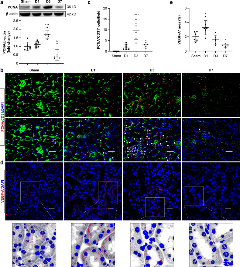
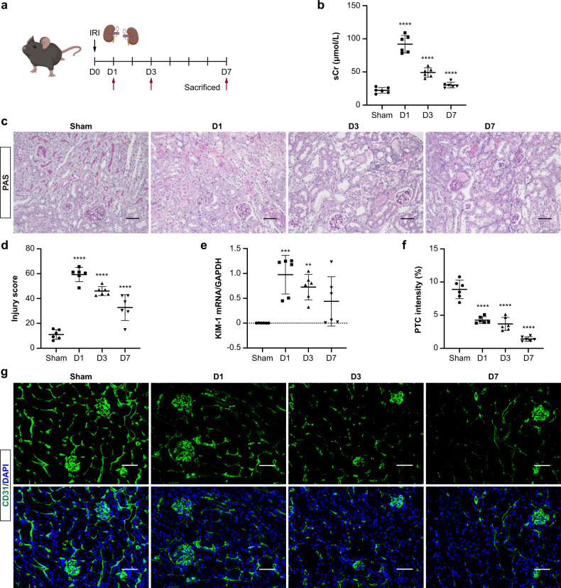
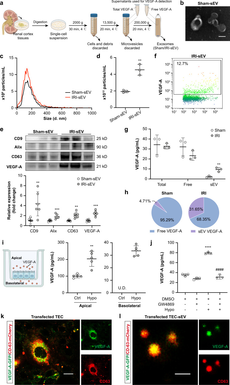
小管周围毛细血管(ptc)在结构和功能上与肾小管密切相关,两者都是急性肾损伤(AKI)发生和进展的关键调节因子。然而,AKI期间ptc和小管相互作用的机制尚不清楚。本研究探讨了AKI后由细胞外小泡(sEV)介导的管-血管串扰的新模式。肾缺血再灌注(I/R)损伤后,ptc内皮细胞增殖增加,血管内皮生长因子- a (VEGF-A)表达增加,细胞质VEGF-A向肾小管细胞基底外侧重新分布。同时,VEGF-A的分泌方式在损伤小管细胞中发生了转变,其通过sEV而非自由形式分泌VEGF-A的倾向要大得多。有趣的是,小管细胞衍生的富含VEGF- a的sEV (sEV-VEGF- a)被证明可以促进内皮细胞的增殖,这是由VEGF受体1和2调节的。此外,通过敲低Rab27a抑制肾sEV分泌,导致体内小管周围内皮细胞的增殖显著减少。重要的是,利用新近认识到的PTC的内源性修复反应,外源性补充VEGF-A + sEV有效地避免了PTC的稀释,改善了肾脏灌注,并阻止了AKI向CKD的过渡。综上所述,我们的研究揭示了肾小管- ptc通过sEV-VEGF-A串扰在AKI后产生的一种新的内在修复反应,这可能是一种有前途的治疗缺血疾病的血管生成策略。
Tubular epithelial cells-derived small extracellular vesicle-VEGF-A promotes peritubular capillary repair in ischemic kidney injury.
Peritubular capillaries (PTCs) are closely related to renal tubules in structure and function, and both are pivotal regulators in the development and progression of acute kidney injury (AKI). However, the mechanisms that underlie the interaction between PTCs and tubules during AKI remain unclear. Here we explored a new mode of tubulovascular crosstalk mediated by small extracellular vesicles (sEV) after AKI. In response to renal ischemia/reperfusion (I/R) injury, endothelial proliferation of PTCs and tubular expression of vascular endothelial growth factor-A (VEGF-A) were increased, accompanied by a remarkable redistribution of cytoplasmic VEGF-A to the basolateral side of tubular cells. Meanwhile, the secretion mode of VEGF-A was converted in the injured tubular cells, which showed a much greater tendency to secrete VEGF-A via sEV other than the free form. Interestingly, tubular cell-derived VEGF-A-enriched sEV (sEV-VEGF-A) turned out to promote endothelial proliferation which was regulated by VEGF receptors 1 and 2. Furthermore, inhibition of renal sEV secretion by Rab27a knockdown resulted in a significant decrease in the proliferation of peritubular endothelial cells in vivo. Importantly, taking advantage of the newly recognized endogenous repair response of PTCs, exogenous supplementation of VEGF-A + sEV efficiently recused PTC rarefaction, improved renal perfusion, and halted the AKI to CKD transition. Taken together, our study uncovered a novel intrinsic repair response after AKI through renal tubule-PTC crosstalk via sEV-VEGF-A, which could be exploited as a promising therapeutic angiogenesis strategy in diseases with ischemia.
期刊介绍:
Regenerative Medicine, an innovative online-only journal, aims to advance research in the field of repairing and regenerating damaged tissues and organs within the human body. As a part of the prestigious Nature Partner Journals series and in partnership with ARMI, this high-quality, open access journal serves as a platform for scientists to explore effective therapies that harness the body's natural regenerative capabilities. With a focus on understanding the fundamental mechanisms of tissue damage and regeneration, npj Regenerative Medicine actively encourages studies that bridge the gap between basic research and clinical tissue repair strategies.


 分享
分享
 求助内容:
求助内容: 应助结果提醒方式:
 扫码关注我们
