Mohamed A M Alsafy, Samir A A El-Gendy, Basma M Kamal, Catrin S Rutland, Hanan H Abd-Elhafeez, Soha Soliman, Ahmed N ELKhamary, Ahmed G Nomir
{"title":"单峰骆驼(Camelus dromedarius)的心脏心室:来自断层解剖、3D计算机断层扫描和形态测量学的新见解。","authors":"Mohamed A M Alsafy, Samir A A El-Gendy, Basma M Kamal, Catrin S Rutland, Hanan H Abd-Elhafeez, Soha Soliman, Ahmed N ELKhamary, Ahmed G Nomir","doi":"10.1186/s40850-023-00173-w","DOIUrl":null,"url":null,"abstract":"<p><strong>Background: </strong>Dromedary camel heart morphology is a crucial research topic with clinical applications. The study aims to understand the dromedary camel anatomy, morphology, and architecture of the ventricular mass.</p><p><strong>Results: </strong>Sagittal and transverse gross sections were compared to sagittal, transverse, and 3D render volume reconstruction computed tomography (CT) scans. The subepicardial fat, which covered the heart base, the coronary groove (sulcus coronarius), the left longitudinal interventricular groove (sulcus interventricularis paraconalis), and the right longitudinal interventricular groove (sulcus interventricularis subsinuosus), had a relatively low density with a homogeneous appearance in the 3D render volume CT. The pericardium in the color cardiac window was identified better than the black and white window (ghost). Transverse and sagittal CT scans demonstrated the internal structures of the heart, including the right atrioventricular orifice (ostium atrioventriculare dextrum), right atrioventricular orifice (ostium atrioventriculare sinistrum), and aortic orifice (ostium aortae), chordae tendineae, the cusps of the valves (cuspis valvae), and the papillary muscles (musculi papillares). The papillary muscle (musculi papillares) was presented with a more moderate density than the rest of the heart, and the cusps of the valves (cuspis valvae) had a lower density. The ventricular wall (margo ventricularis) exhibited different densities: the outer part was hyperdense, while the inner part was hypodense. The thicknesses of the ventricular mural wall and the interventricular septum (septum atrioventriculare) were highest at the midpoint of the ventricular mass, and the lowest value was present toward the apical part. The coronary groove (sulcus coronarius) circumference measured 51.14 ± 0.72 cm, and the fat in the coronary groove (sulcus coronarius) (56 ± 6.55 cm<sup>2</sup>) represented 28.7% of the total cross-sectional area.</p><p><strong>Conclusion: </strong>The current study provided more information about ventricular mass measurements by gross and CT analysis on the heart, which provides a valuable guide for future cardiac CT investigations in camels in vivo.</p>","PeriodicalId":48590,"journal":{"name":"BMC Zoology","volume":"8 1","pages":"12"},"PeriodicalIF":1.7000,"publicationDate":"2023-08-18","publicationTypes":"Journal Article","fieldsOfStudy":null,"isOpenAccess":false,"openAccessPdf":"https://www.ncbi.nlm.nih.gov/pmc/articles/PMC10436409/pdf/","citationCount":"0","resultStr":"{\"title\":\"Heart ventricles of the dromedary camel (Camelus dromedarius): new insights from sectional anatomy, 3D computed tomography, and morphometry.\",\"authors\":\"Mohamed A M Alsafy, Samir A A El-Gendy, Basma M Kamal, Catrin S Rutland, Hanan H Abd-Elhafeez, Soha Soliman, Ahmed N ELKhamary, Ahmed G Nomir\",\"doi\":\"10.1186/s40850-023-00173-w\",\"DOIUrl\":null,\"url\":null,\"abstract\":\"<p><strong>Background: </strong>Dromedary camel heart morphology is a crucial research topic with clinical applications. The study aims to understand the dromedary camel anatomy, morphology, and architecture of the ventricular mass.</p><p><strong>Results: </strong>Sagittal and transverse gross sections were compared to sagittal, transverse, and 3D render volume reconstruction computed tomography (CT) scans. The subepicardial fat, which covered the heart base, the coronary groove (sulcus coronarius), the left longitudinal interventricular groove (sulcus interventricularis paraconalis), and the right longitudinal interventricular groove (sulcus interventricularis subsinuosus), had a relatively low density with a homogeneous appearance in the 3D render volume CT. The pericardium in the color cardiac window was identified better than the black and white window (ghost). Transverse and sagittal CT scans demonstrated the internal structures of the heart, including the right atrioventricular orifice (ostium atrioventriculare dextrum), right atrioventricular orifice (ostium atrioventriculare sinistrum), and aortic orifice (ostium aortae), chordae tendineae, the cusps of the valves (cuspis valvae), and the papillary muscles (musculi papillares). The papillary muscle (musculi papillares) was presented with a more moderate density than the rest of the heart, and the cusps of the valves (cuspis valvae) had a lower density. The ventricular wall (margo ventricularis) exhibited different densities: the outer part was hyperdense, while the inner part was hypodense. The thicknesses of the ventricular mural wall and the interventricular septum (septum atrioventriculare) were highest at the midpoint of the ventricular mass, and the lowest value was present toward the apical part. The coronary groove (sulcus coronarius) circumference measured 51.14 ± 0.72 cm, and the fat in the coronary groove (sulcus coronarius) (56 ± 6.55 cm<sup>2</sup>) represented 28.7% of the total cross-sectional area.</p><p><strong>Conclusion: </strong>The current study provided more information about ventricular mass measurements by gross and CT analysis on the heart, which provides a valuable guide for future cardiac CT investigations in camels in vivo.</p>\",\"PeriodicalId\":48590,\"journal\":{\"name\":\"BMC Zoology\",\"volume\":\"8 1\",\"pages\":\"12\"},\"PeriodicalIF\":1.7000,\"publicationDate\":\"2023-08-18\",\"publicationTypes\":\"Journal Article\",\"fieldsOfStudy\":null,\"isOpenAccess\":false,\"openAccessPdf\":\"https://www.ncbi.nlm.nih.gov/pmc/articles/PMC10436409/pdf/\",\"citationCount\":\"0\",\"resultStr\":null,\"platform\":\"Semanticscholar\",\"paperid\":null,\"PeriodicalName\":\"BMC Zoology\",\"FirstCategoryId\":\"99\",\"ListUrlMain\":\"https://doi.org/10.1186/s40850-023-00173-w\",\"RegionNum\":3,\"RegionCategory\":\"生物学\",\"ArticlePicture\":[],\"TitleCN\":null,\"AbstractTextCN\":null,\"PMCID\":null,\"EPubDate\":\"\",\"PubModel\":\"\",\"JCR\":\"Q2\",\"JCRName\":\"ZOOLOGY\",\"Score\":null,\"Total\":0}","platform":"Semanticscholar","paperid":null,"PeriodicalName":"BMC Zoology","FirstCategoryId":"99","ListUrlMain":"https://doi.org/10.1186/s40850-023-00173-w","RegionNum":3,"RegionCategory":"生物学","ArticlePicture":[],"TitleCN":null,"AbstractTextCN":null,"PMCID":null,"EPubDate":"","PubModel":"","JCR":"Q2","JCRName":"ZOOLOGY","Score":null,"Total":0}
Heart ventricles of the dromedary camel (Camelus dromedarius): new insights from sectional anatomy, 3D computed tomography, and morphometry.
Background: Dromedary camel heart morphology is a crucial research topic with clinical applications. The study aims to understand the dromedary camel anatomy, morphology, and architecture of the ventricular mass.
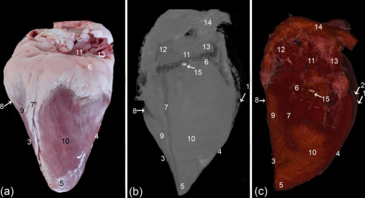
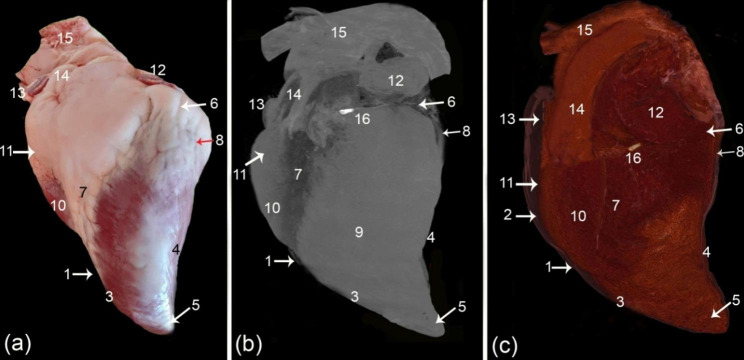
Results: Sagittal and transverse gross sections were compared to sagittal, transverse, and 3D render volume reconstruction computed tomography (CT) scans. The subepicardial fat, which covered the heart base, the coronary groove (sulcus coronarius), the left longitudinal interventricular groove (sulcus interventricularis paraconalis), and the right longitudinal interventricular groove (sulcus interventricularis subsinuosus), had a relatively low density with a homogeneous appearance in the 3D render volume CT. The pericardium in the color cardiac window was identified better than the black and white window (ghost). Transverse and sagittal CT scans demonstrated the internal structures of the heart, including the right atrioventricular orifice (ostium atrioventriculare dextrum), right atrioventricular orifice (ostium atrioventriculare sinistrum), and aortic orifice (ostium aortae), chordae tendineae, the cusps of the valves (cuspis valvae), and the papillary muscles (musculi papillares). The papillary muscle (musculi papillares) was presented with a more moderate density than the rest of the heart, and the cusps of the valves (cuspis valvae) had a lower density. The ventricular wall (margo ventricularis) exhibited different densities: the outer part was hyperdense, while the inner part was hypodense. The thicknesses of the ventricular mural wall and the interventricular septum (septum atrioventriculare) were highest at the midpoint of the ventricular mass, and the lowest value was present toward the apical part. The coronary groove (sulcus coronarius) circumference measured 51.14 ± 0.72 cm, and the fat in the coronary groove (sulcus coronarius) (56 ± 6.55 cm2) represented 28.7% of the total cross-sectional area.
Conclusion: The current study provided more information about ventricular mass measurements by gross and CT analysis on the heart, which provides a valuable guide for future cardiac CT investigations in camels in vivo.
BMC ZoologyAgricultural and Biological Sciences-Animal Science and Zoology
CiteScore
2.30
自引率
6.20%
发文量
53
审稿时长
24 weeks
期刊介绍:
BMC Zoology is an open access, peer-reviewed journal that considers articles on all aspects of zoology, including physiology, mechanistic and functional studies, anatomy, life history, behavior, signalling and communication, cognition, parasitism, taxonomy and conservation.


 分享
分享
 求助内容:
求助内容: 应助结果提醒方式:
 扫码关注我们
