{"title":"正颌手术后意外颞下颌关节盘复位:1例长期影像学随访报告。","authors":"Hak-Sun Kim, Sang-Sun Han, Chena Lee","doi":"10.5624/isd.20220048","DOIUrl":null,"url":null,"abstract":"<p><p>This report presents a rare case where a displaced temporomandibular joint (TMJ) disc was reduced to its normal position after orthognathic surgery, and long-term magnetic resonance imaging (MRI) follow-up visualized these postoperative changes. A 22-year-old male patient presented for facial asymmetry. He also complained of pain in the right TMJ area, and MRI showed disc displacements in both TMJs. After orthognathic surgery for facial asymmetry correction, the TMJ was re-evaluated. The symptom had resolved and the disc was positioned within the normal range during mouth opening. However, 6 and a half years after surgery, he complained of recurrent pain in the right joint, and MRI revealed medial disc displacement in the right TMJ. In conclusion, the influence of orthognathic surgery on the disc position might continue for a long time until the TMJ adapts to the new position. Careful and long-term follow-up is suggested to assess the TMJ complex.</p>","PeriodicalId":51714,"journal":{"name":"Imaging Science in Dentistry","volume":"52 4","pages":"409-413"},"PeriodicalIF":1.7000,"publicationDate":"2022-12-01","publicationTypes":"Journal Article","fieldsOfStudy":null,"isOpenAccess":false,"openAccessPdf":"https://ftp.ncbi.nlm.nih.gov/pub/pmc/oa_pdf/e4/94/isd-52-409.PMC9807793.pdf","citationCount":"0","resultStr":"{\"title\":\"Unintentional temporomandibular joint disc reduction after orthognathic surgery: A case report with long-term imaging follow-up.\",\"authors\":\"Hak-Sun Kim, Sang-Sun Han, Chena Lee\",\"doi\":\"10.5624/isd.20220048\",\"DOIUrl\":null,\"url\":null,\"abstract\":\"<p><p>This report presents a rare case where a displaced temporomandibular joint (TMJ) disc was reduced to its normal position after orthognathic surgery, and long-term magnetic resonance imaging (MRI) follow-up visualized these postoperative changes. A 22-year-old male patient presented for facial asymmetry. He also complained of pain in the right TMJ area, and MRI showed disc displacements in both TMJs. After orthognathic surgery for facial asymmetry correction, the TMJ was re-evaluated. The symptom had resolved and the disc was positioned within the normal range during mouth opening. However, 6 and a half years after surgery, he complained of recurrent pain in the right joint, and MRI revealed medial disc displacement in the right TMJ. In conclusion, the influence of orthognathic surgery on the disc position might continue for a long time until the TMJ adapts to the new position. Careful and long-term follow-up is suggested to assess the TMJ complex.</p>\",\"PeriodicalId\":51714,\"journal\":{\"name\":\"Imaging Science in Dentistry\",\"volume\":\"52 4\",\"pages\":\"409-413\"},\"PeriodicalIF\":1.7000,\"publicationDate\":\"2022-12-01\",\"publicationTypes\":\"Journal Article\",\"fieldsOfStudy\":null,\"isOpenAccess\":false,\"openAccessPdf\":\"https://ftp.ncbi.nlm.nih.gov/pub/pmc/oa_pdf/e4/94/isd-52-409.PMC9807793.pdf\",\"citationCount\":\"0\",\"resultStr\":null,\"platform\":\"Semanticscholar\",\"paperid\":null,\"PeriodicalName\":\"Imaging Science in Dentistry\",\"FirstCategoryId\":\"1085\",\"ListUrlMain\":\"https://doi.org/10.5624/isd.20220048\",\"RegionNum\":0,\"RegionCategory\":null,\"ArticlePicture\":[],\"TitleCN\":null,\"AbstractTextCN\":null,\"PMCID\":null,\"EPubDate\":\"\",\"PubModel\":\"\",\"JCR\":\"Q3\",\"JCRName\":\"DENTISTRY, ORAL SURGERY & MEDICINE\",\"Score\":null,\"Total\":0}","platform":"Semanticscholar","paperid":null,"PeriodicalName":"Imaging Science in Dentistry","FirstCategoryId":"1085","ListUrlMain":"https://doi.org/10.5624/isd.20220048","RegionNum":0,"RegionCategory":null,"ArticlePicture":[],"TitleCN":null,"AbstractTextCN":null,"PMCID":null,"EPubDate":"","PubModel":"","JCR":"Q3","JCRName":"DENTISTRY, ORAL SURGERY & MEDICINE","Score":null,"Total":0}
Unintentional temporomandibular joint disc reduction after orthognathic surgery: A case report with long-term imaging follow-up.
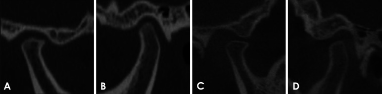
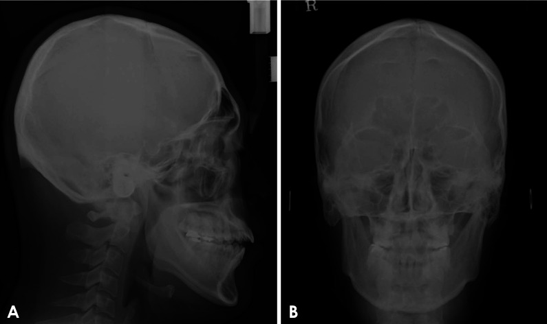
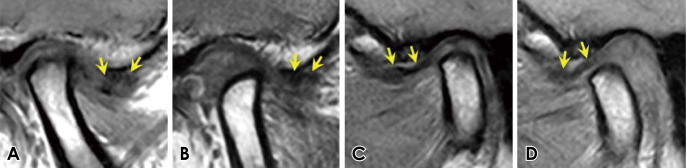
This report presents a rare case where a displaced temporomandibular joint (TMJ) disc was reduced to its normal position after orthognathic surgery, and long-term magnetic resonance imaging (MRI) follow-up visualized these postoperative changes. A 22-year-old male patient presented for facial asymmetry. He also complained of pain in the right TMJ area, and MRI showed disc displacements in both TMJs. After orthognathic surgery for facial asymmetry correction, the TMJ was re-evaluated. The symptom had resolved and the disc was positioned within the normal range during mouth opening. However, 6 and a half years after surgery, he complained of recurrent pain in the right joint, and MRI revealed medial disc displacement in the right TMJ. In conclusion, the influence of orthognathic surgery on the disc position might continue for a long time until the TMJ adapts to the new position. Careful and long-term follow-up is suggested to assess the TMJ complex.


 分享
分享
 求助内容:
求助内容: 应助结果提醒方式:
 扫码关注我们
